
The Anterolateral Thigh (ALT) Flap: Comprehensive Surgical Guide for Design, Harvest, Donor-Site Closure, and Inset in Partial Glossectomy Reconstruction
Authors: Thomas S. Lee, MD; Eli Gordin, MD; Chris Kandl, MD
Published: 1/20/2026
The anterolateral thigh (ALT) flap is a workhorse in head and neck reconstruction owing to its long pedicle, adjustable thickness, option for sensate harvest, and low donor-site morbidity. This manuscript accompanies Dr. Lee’s surgical video and follows his operative sequence while integrating pearls from Drs. Gordin and Kandl, as well as evidence from large clinical series and cadaveric studies [2–11]. We provide a practical, literature-aligned, stepwise guide for flap design, harvest, donor-site closure, and inset for partial glossectomy.
2.1 Patient Positioning and the AP Line
Position the patient supine with the patella oriented straight upward to avoid external rotation that would shift perforator projection on the skin. A stack of towels can be placed beneath the ipsilateral hip to maintain neutral alignment. Dr. Yu recommends taping the feet together to prevent lateral rotation [3].
Draw a straight line from the anterior superior iliac spine (ASIS) to the superolateral corner of the patella (the AP line); this serves as the reference axis for perforator prediction [3]. You can also attempt to palpate for the groove between the rectus femoris and vatus lateralis muscles.
2.2 ABC Perforator System
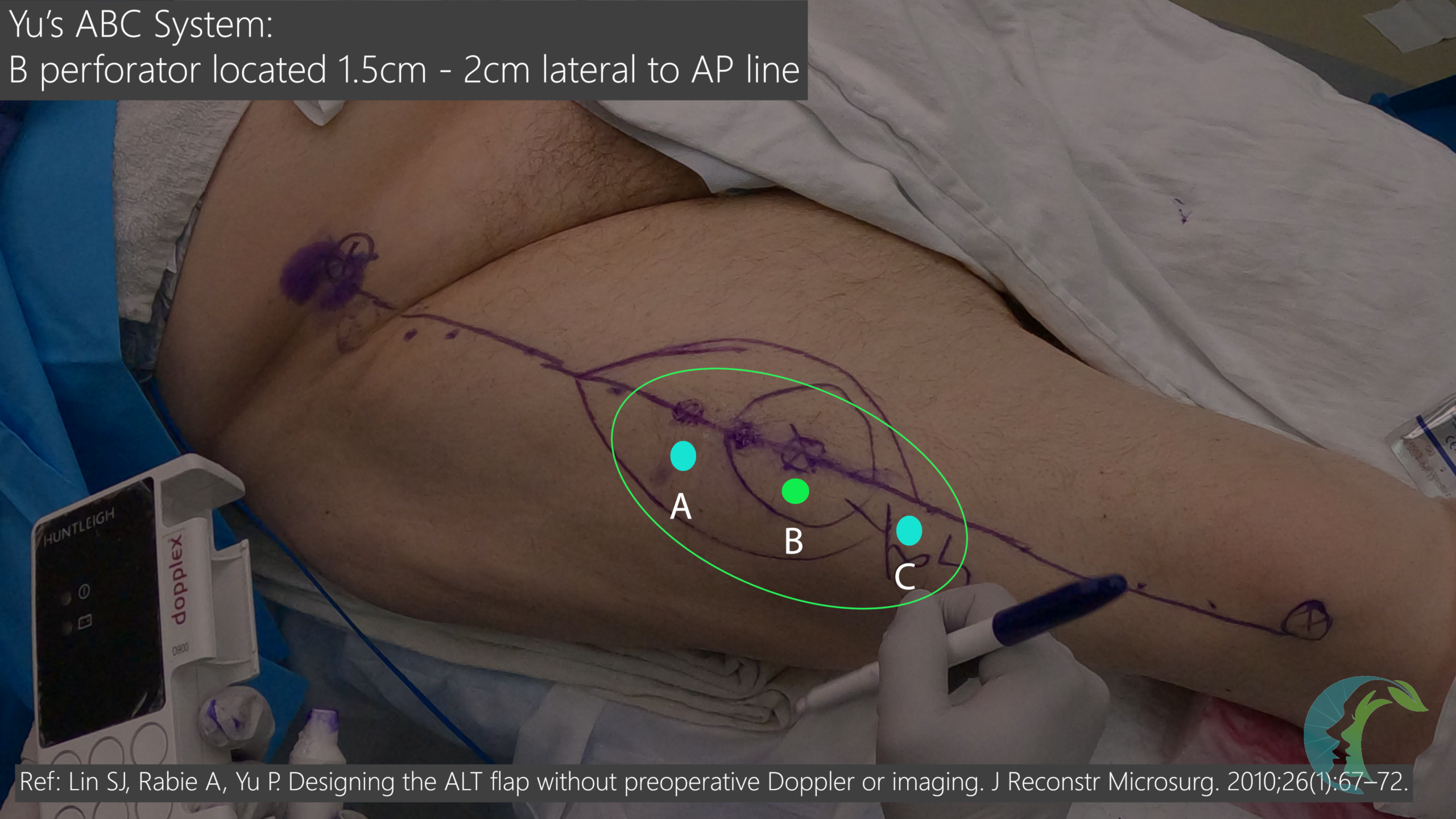
In an updated study, Lin, Rabie and Yu described a reproducible map for three reliable perforator sites (A, B, C) along the ASIS–lateral patella line. The B point lies at the midpoint, approximately 1.5 cm lateral to the AP line. The A point is 5 cm proximal to B; the C point is 5 cm distal to B.
In a series of 124 flaps designed without Doppler using this system, intraoperative locations corresponded closely to predictions. Western series demonstrate similar spacing, with the B perforator included in ~87–93% of viable ALT flaps. The A perforator is present in ~49–53% and the C perforator in ~59–63% [3,4]. This supports designing the skin paddle around the B perforator whenever feasible.
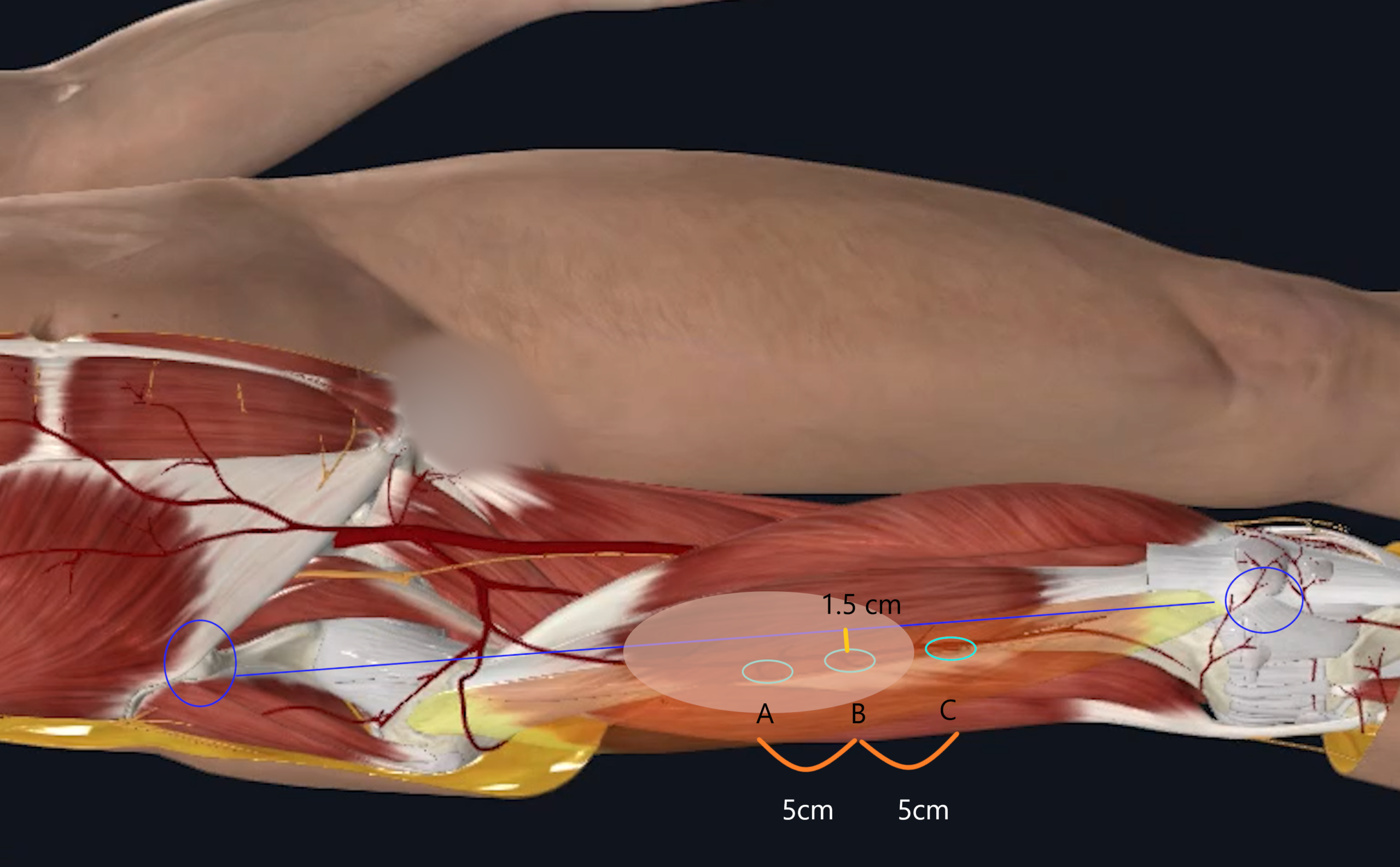 Figure 1. Right Leg. AP line connects anterior superior iliac spine and lateral patella. B point is located at its halfway point and 1.5 – 2cm lateral to the AP line. There are usually 3 perforators at A, B & C. A point is 5cm proximal and C point is 5cm distal to the B point. B point perforator is most commonly included in 90% of ALT flaps. Adapted from Reference 3 (Lin et al).
2.3 Perforator Types (Septocutaneous vs Musculocutaneous)
In Wei’s experience with 672 ALT flaps, cutaneous vessels were predominantly musculocutaneous (~87.1%) and less commonly septocutaneous (~12.9%). Both courses are reliable when dissected retrograde to the descending branch of the lateral circumflex femoral artery (DB‑LCFA) [2].
2.4 Perforator Origin Types (Yu Classification)
Three perforator origin patterns are clinically relevant. Correctly identifying the source vessel is critical to a successful harvest.
Type I, present in ~90% of cases, features perforators arising from the DB‑LCFA. In the remaining ~10%, variations may complicate ALT use.
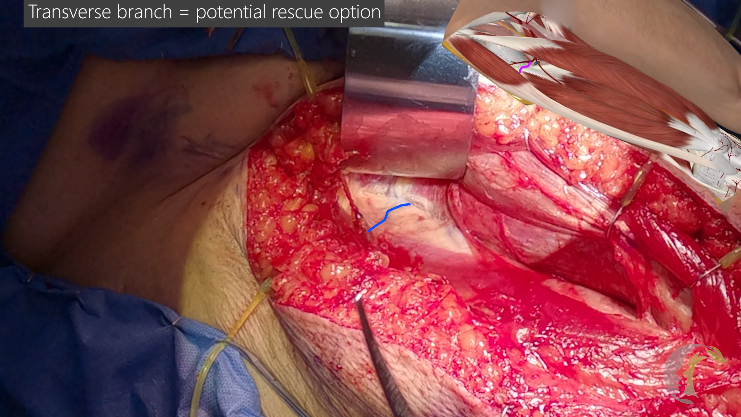
In Type II, the B perforator arises from the transverse branch; in these cases, the flap should be based to include the transverse branch. Type II occurred in ~4%.
In Type III, the B perforator originates from the profunda femoris. These perforators tend to be of small caliber with short pedicles and are typically unsuitable for free transfer; Yu recommends abandoning the ipsilateral ALT Type III and harvesting from the contralateral thigh instead [4]. In all cases, confirm that the perforator traces back to the DB‑LCFA or the transverse branch and adjust flap design accordingly.
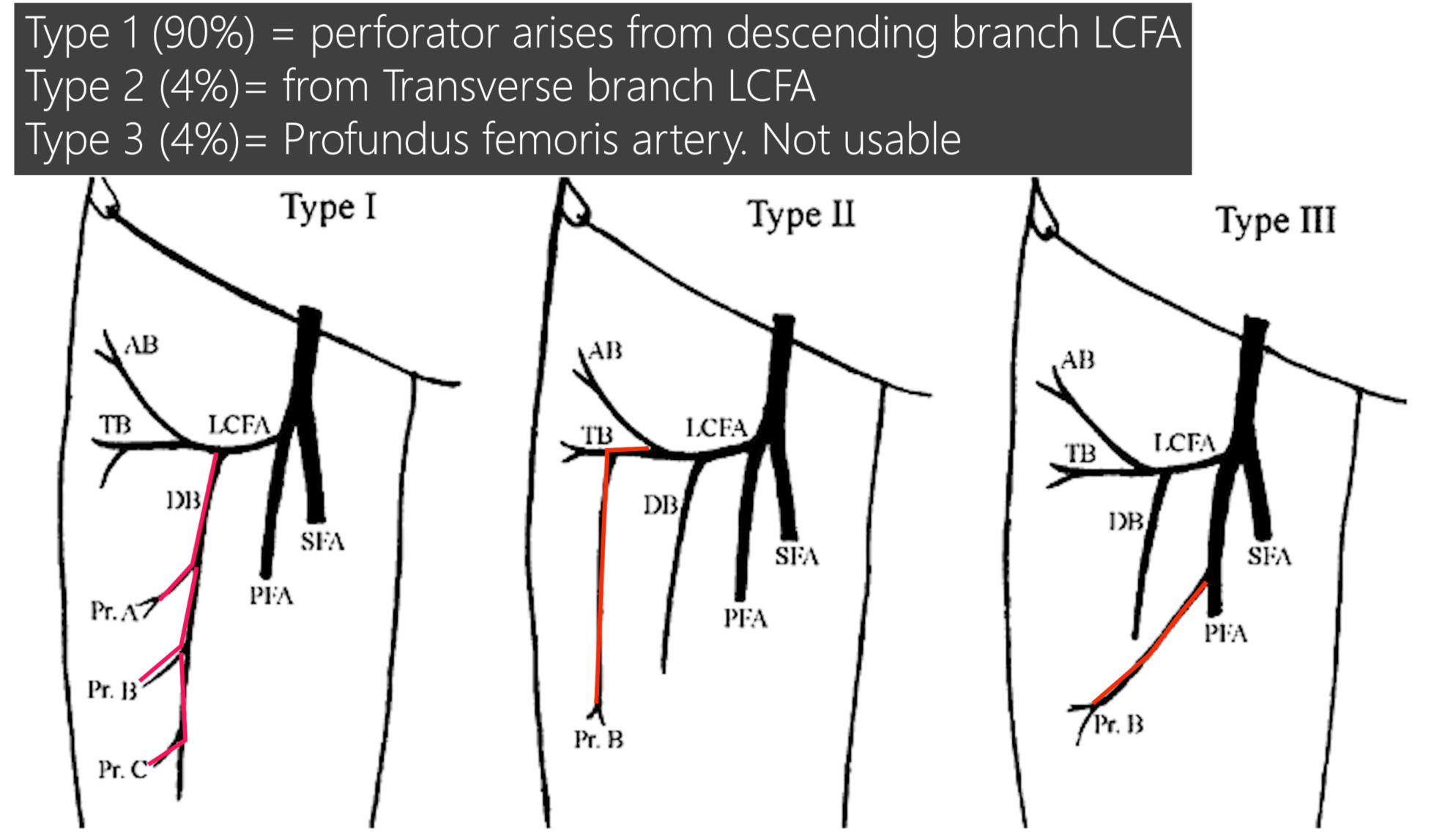
Figure 2. Three types of anatomic variations of the anterolateral thigh (ALT) flap pedicle. In type I, cutaneous perforators (Pr. A, B, and C) originate from the descending branch (DB). In type II, the cutaneous perforator (Pr. B) originates from the transverse branch (TB). In type III, the cutaneous perforator (Pr. B) originates from the profundus femoris artery (PFA) directly. SFA,superficial femoral artery; LCFA, lateral circumflex femoris artery; AB, ascending branch. From reference 4 (Yu).
2.5 Western Thickness Planning
ALT thickness in Western patients correlates with BMI. At the B level, mean thickness is ~13 mm in men and ~20 mm in women; the A level is ~20% thicker than B, and the C level is ~18% thinner than B. Select the C level when thinner tissue is required for intraoral lining, and the A level when greater bulk is desired (e.g., total glossectomy or external contour) [4]. ALTskin flap designed closer to the knee (near C point) will have greater flap vessel reach and may serve as another important decision factor in flap design.
3.1 Skin Flap Design & Anterior Incision First
3.2 Subfascial Dissection
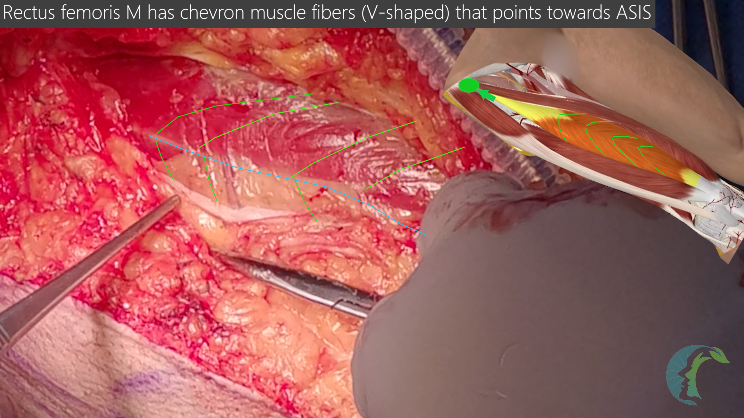
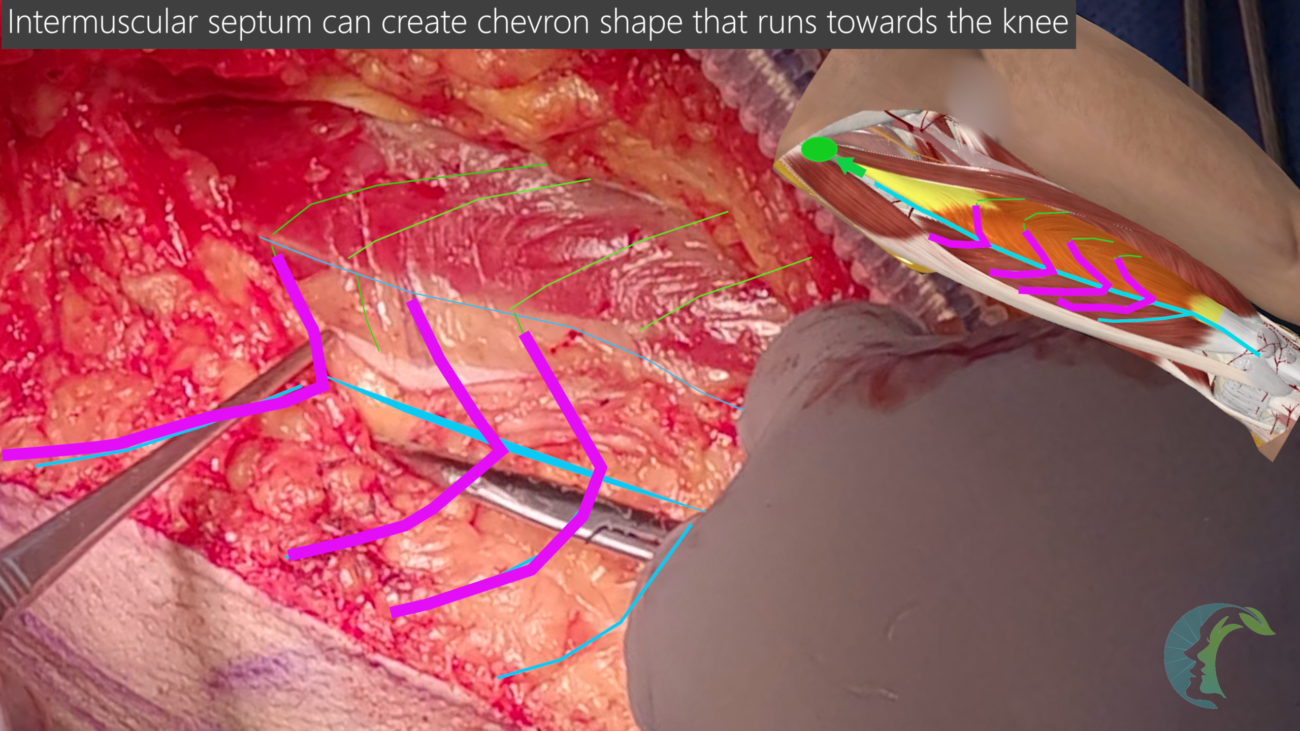
3.3 Identifying the Rectus femoris muscle & vastus lateralis muscle
3.3 Perforator-First Retrograde Dissection
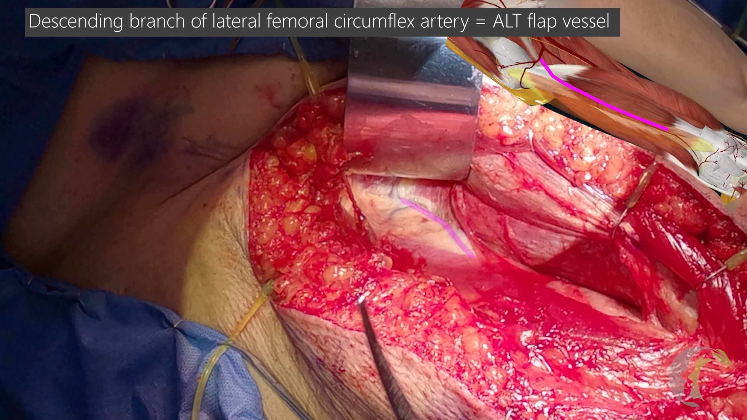
3.4 DB‑LCFA Pedicle Dissection
3.5 Motor Nerve to the Vastus Lateralis (MNVL):
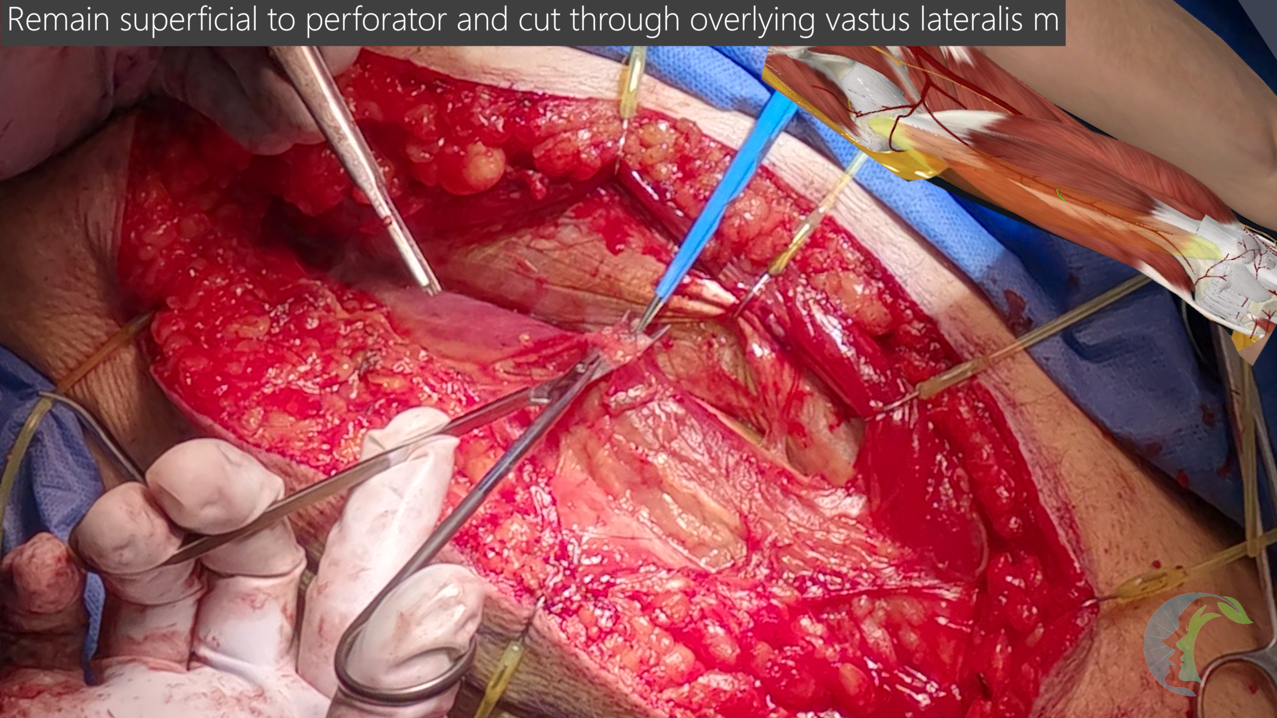
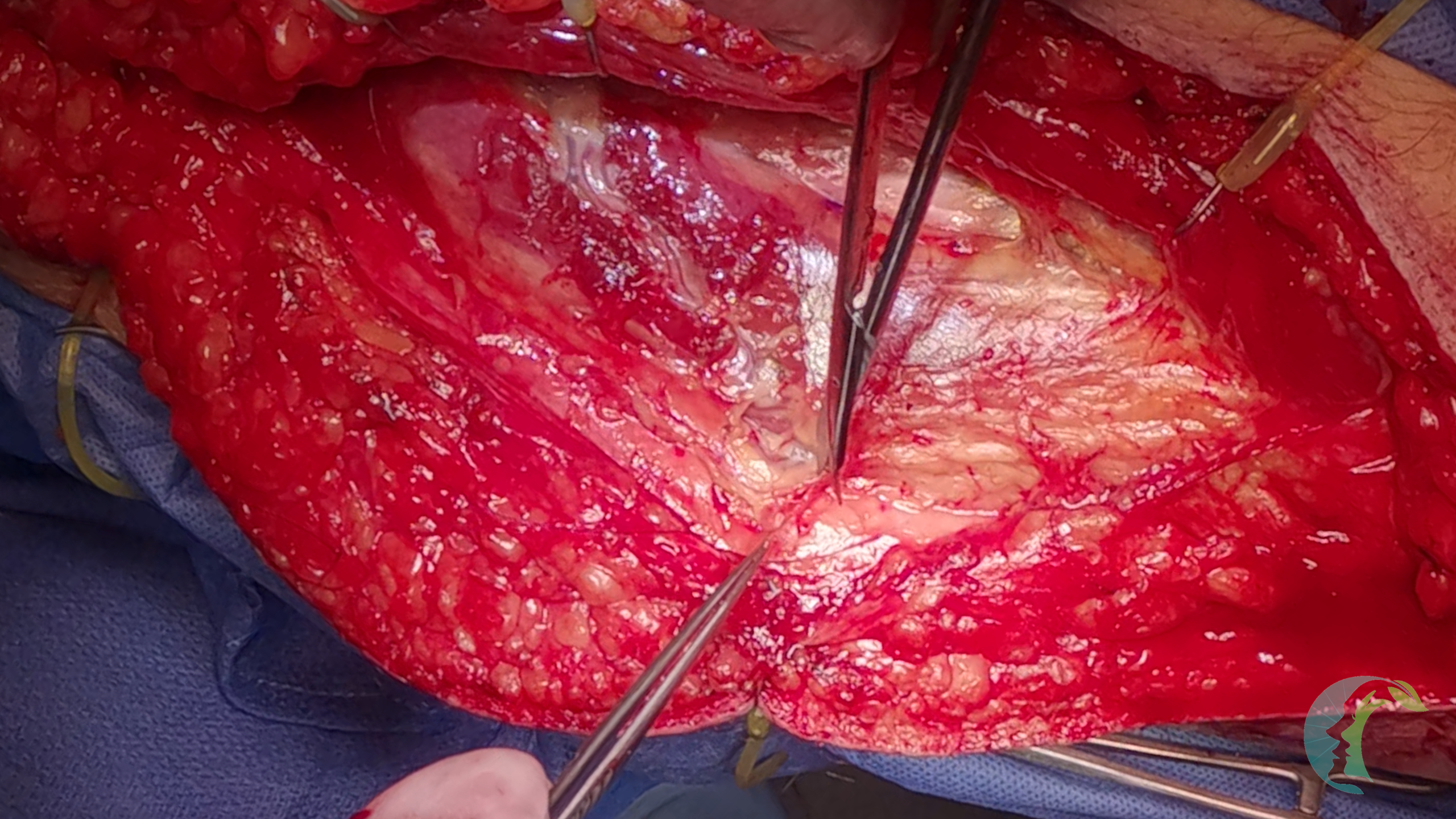
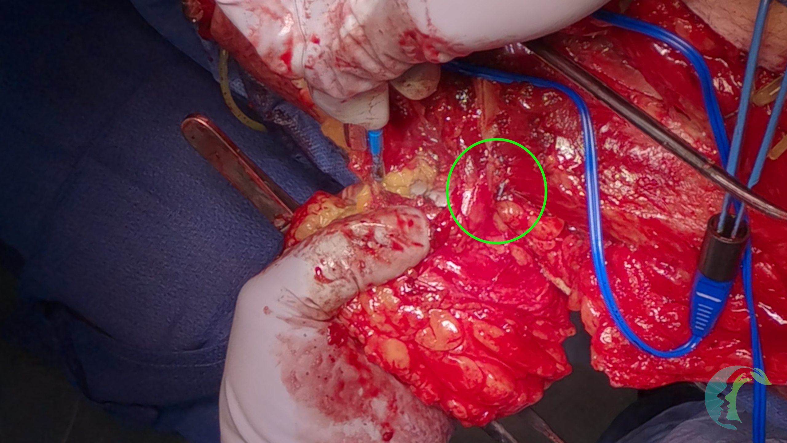
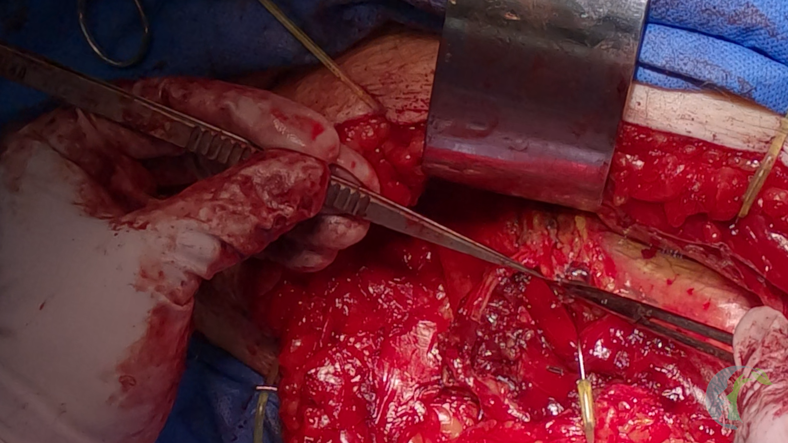
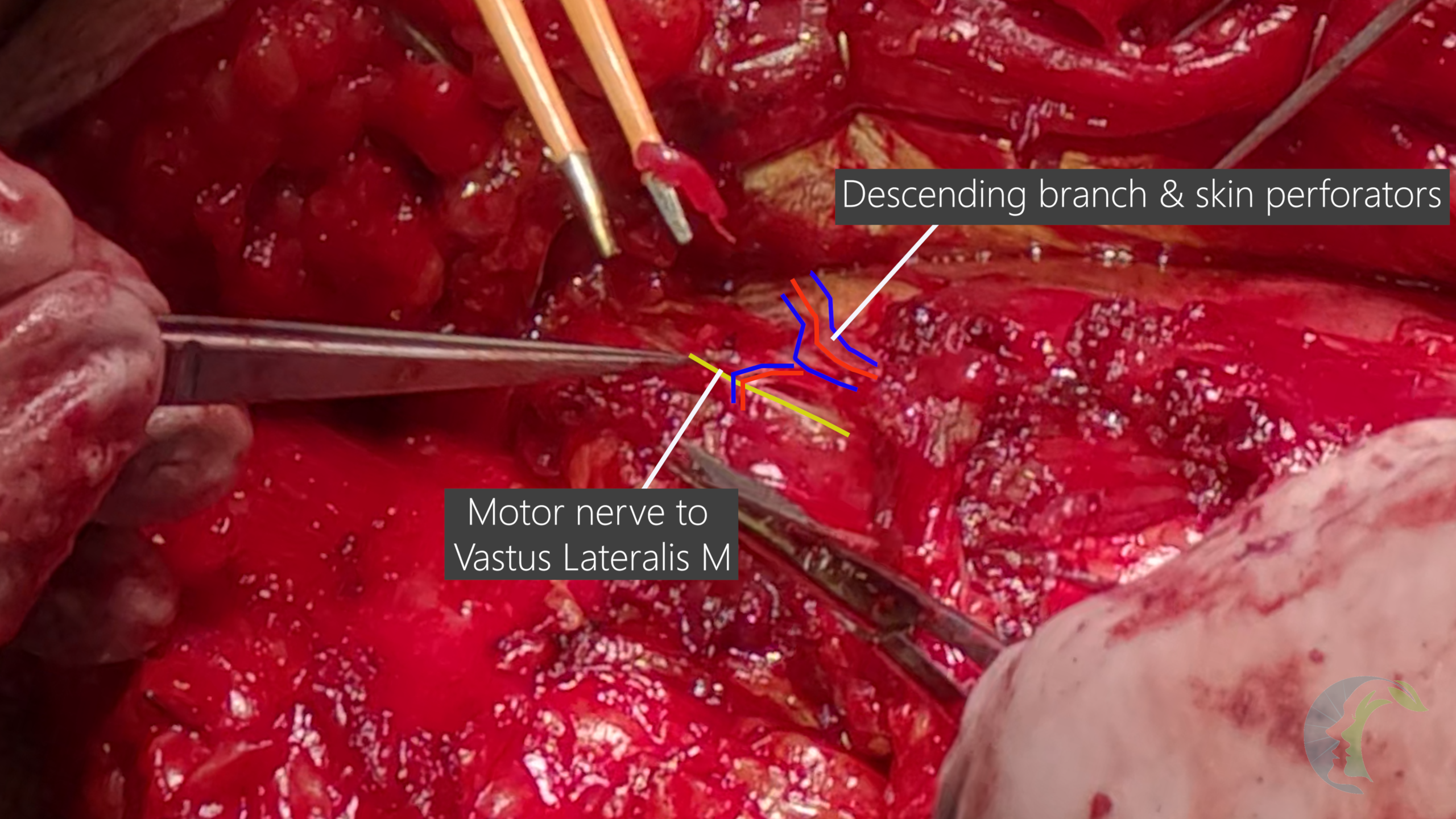
Motor nerve to the vastus lateralis is in close proximity to the flap vessels and its relationship to the vessels can vary greatly. Rozen described three problematic relationships between the MNVL and the vascular pedicle: (1) a motor branch traversing the main pedicle; (2) a branch crossing a perforator ; and (3) a branch passing between adjacent perforators—collectively seen in ~28% of thighs [7]. Casey reported that ~7% of clinical cases required major modification due to nerve position [8]. Revenaugh’s cadaveric study confirmed 3–6 primary branches with extensive arborization and variable relationships to the DB‑LCFA and perforators, supporting selective sacrifice when necessary [9].
Functionally, selective division of a VL motor branch has not been associated with persistent donor weakness; prospective series demonstrate that subjective weakness resolves by approximately 6 months [10]. Similarly, VL muscle harvest has not produced clinically significant long-term dysfunction [10]. Collectively, these studies highlight the importance of prioritizing preservation of flap vasculature; selective sacrifice of the VL motor nerve is acceptable when required, as it is rarely associated with meaningful postoperative donor-site morbidity. With that said, motor branches to other quadriceps muscle should be preserved.
3.6 Thinning and Shaping
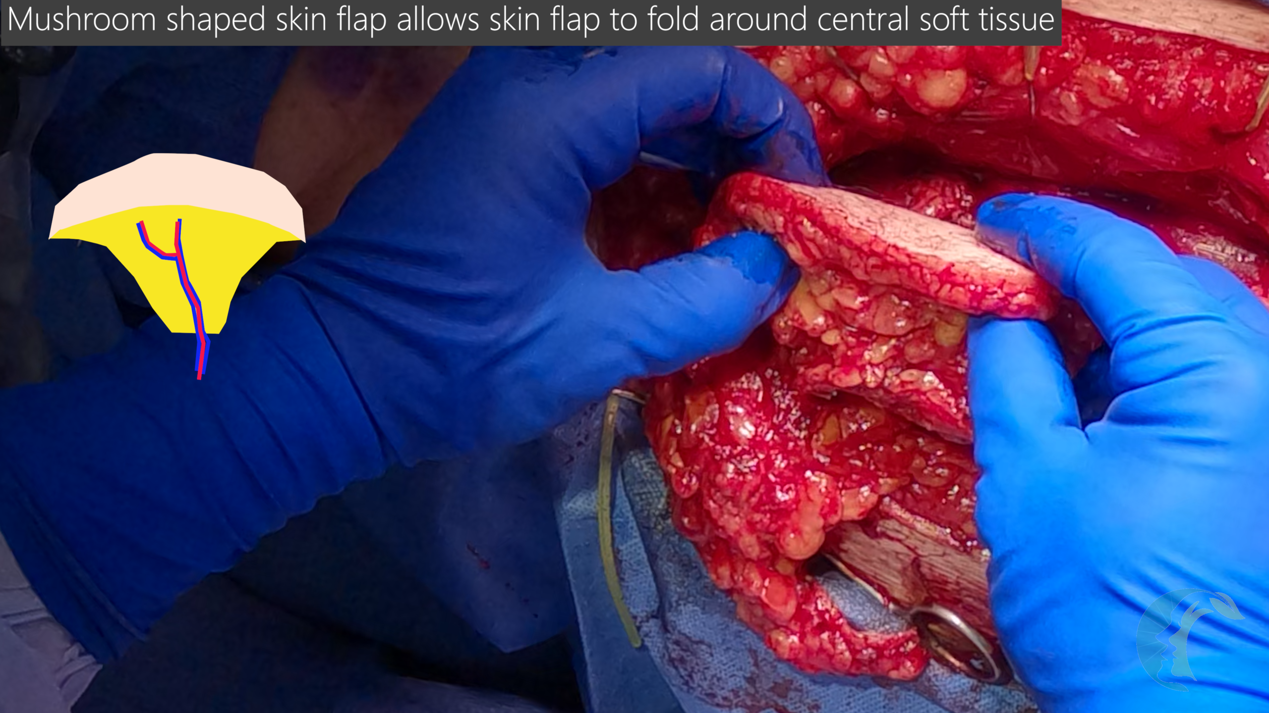
Before harvesting the flap, debulk the flap if necessary by tapering subcutaneous fat while preserving the subdermal plexus (leave 1–2 mm) and protecting the perforator. In thick thighs, the perforator can be dissected 1–2 cm distally into the fat to facilitate safer debulking without compromising inflow [5]. Select the A/B/C level to match thickness requirements [4,5].
3.7 Flap harvest
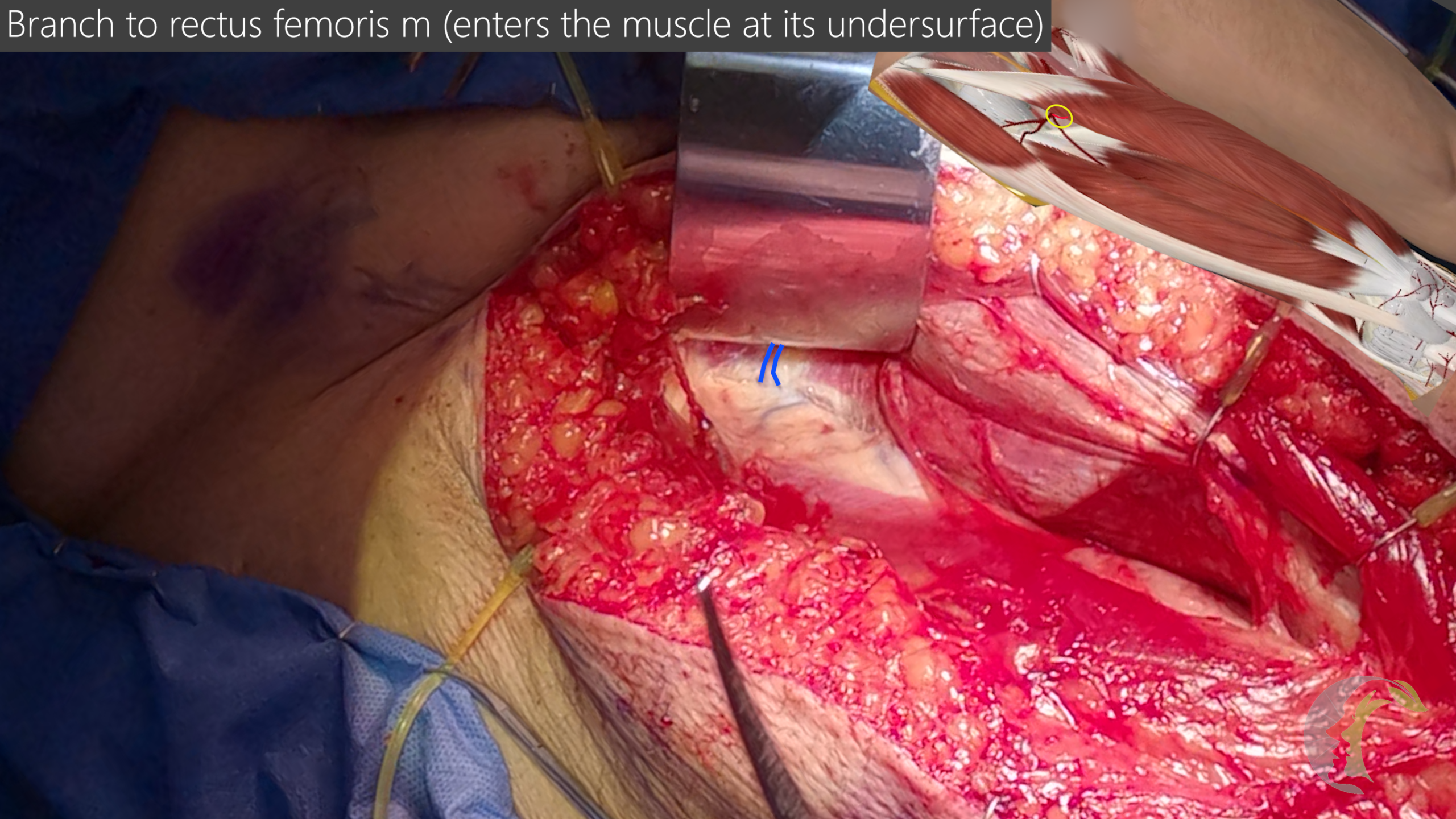
Once head & neck site is ready and the neck vessels have been identified, harvest the ALT flap. The flap vessels are harvested distal to the branch to RF.
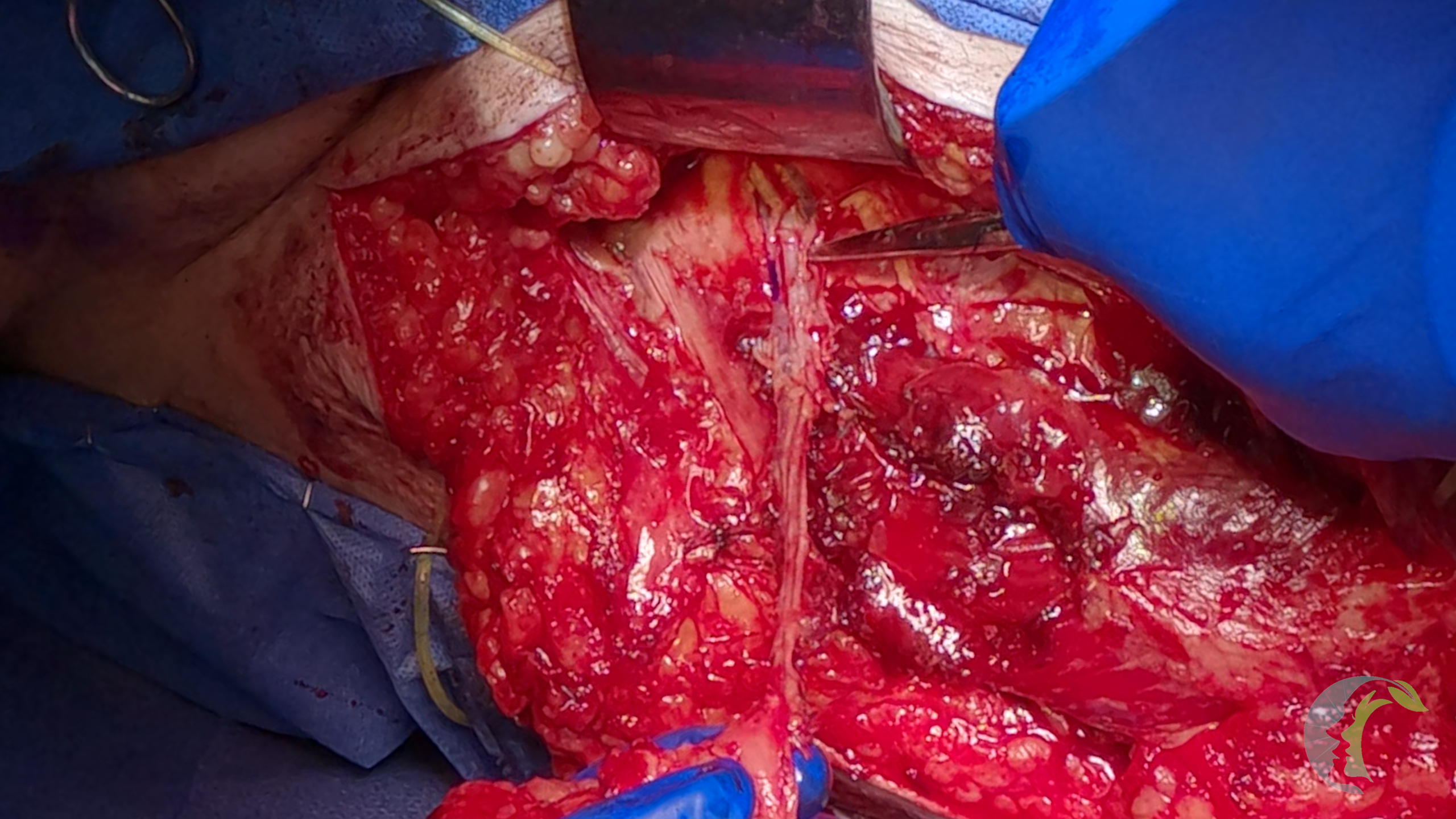
Flap is irrigated with heparinized saline.
4.1 Primary Closure Feasibility
Primary closure depends on flap width, tissue laxity, BMI, and proximity to the knee; there is no universal cutoff. Yu reported primary closure in ~94% of cases in a Western cohort [4].
4.2 When Skin Grafting Is Required
Kimata correlated greater morbidity (gait limitation/knee restriction) with grafted donor sites, wider flaps, and designs closer to the knee. Anticipate increased aftercare if grafting is unavoidable and, when feasible, avoid designing the skin paddle close to the knee [11]. In some reconstructions, however, a more distal design may provide additional pedicle reach and can be appropriate depending on recipient site needs. [1]
If skin grafting is required, the senior authors (Lee and Kandl) prefer a split-thickness skin graft (STSG) harvested from the ipsilateral thigh, secured with negative-pressure wound therapy (NPWT). A critical step is ensuring an airtight seal at the graft site to prevent loss of vacuum and subsequent Jackson-Pratt (JP) drain leakage. Following maximal primary closure of the leg incision, attention is directed to the graft recipient site. A circumferential, running, buried Vicryl suture is placed through the subdermal layer and secured to the underlying muscular wound bed. The STSG is then applied and the NPWT dressing is maintained for seven days.
4.3 Seroma Prevention and Aftercare
Besides using drain suction, Dr. Gordin recommends applying early compression (ACE wrap) [5]. Needle aspiration may be required for seroma. In Hanasono’s prospective series of 220 ALT flaps, reported complications were: seroma 5%, dehiscence 2%, hematoma 1%, and infection 1%—overall low rates [10].
4.4 Muscle and Nerve Handling
Avoid forced muscle closure that may increase compartment pressures although this is very rare. Re-approximate small VL cuffs when feasible. Expect lateral femoral cutaneous nerve (LFCN) numbness postoperatively; clinically significant donor extremity motor dysfunction is rare [1,10].
5.1 Goals
Recreate functional tongue bulk (anticipate ~50% volume loss after radiation therapy) and achieve a watertight closure to protect the neck from salivary contamination and fistula formation. The reconstructed tongue bulk should ideally allow the patient to contact the palate to optimize swallowing function [1].
5.2 Pedicle Routing and Protection
Before you can inset the flap, you should pass the flap vessels into the neck first.
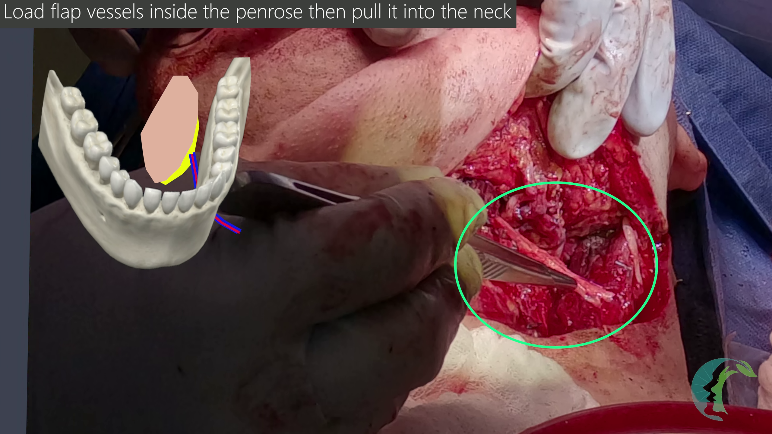
5.3 Posterior Closure First (Pre-reperfusion)
Complete inset along the deep and posterior aspects of the defect before reperfusion (ideally within 1.5–2 hours of ischemia). This avoids edema-limited visualization and facilitates precise suturing [1].
5.4 Circumdental Suspension
When securing skin flap along the lingual gingiva, use circumdental sutures that wrap around teeth to resist gravitational descent of the skin flap which may lead to salivary fistula [1].
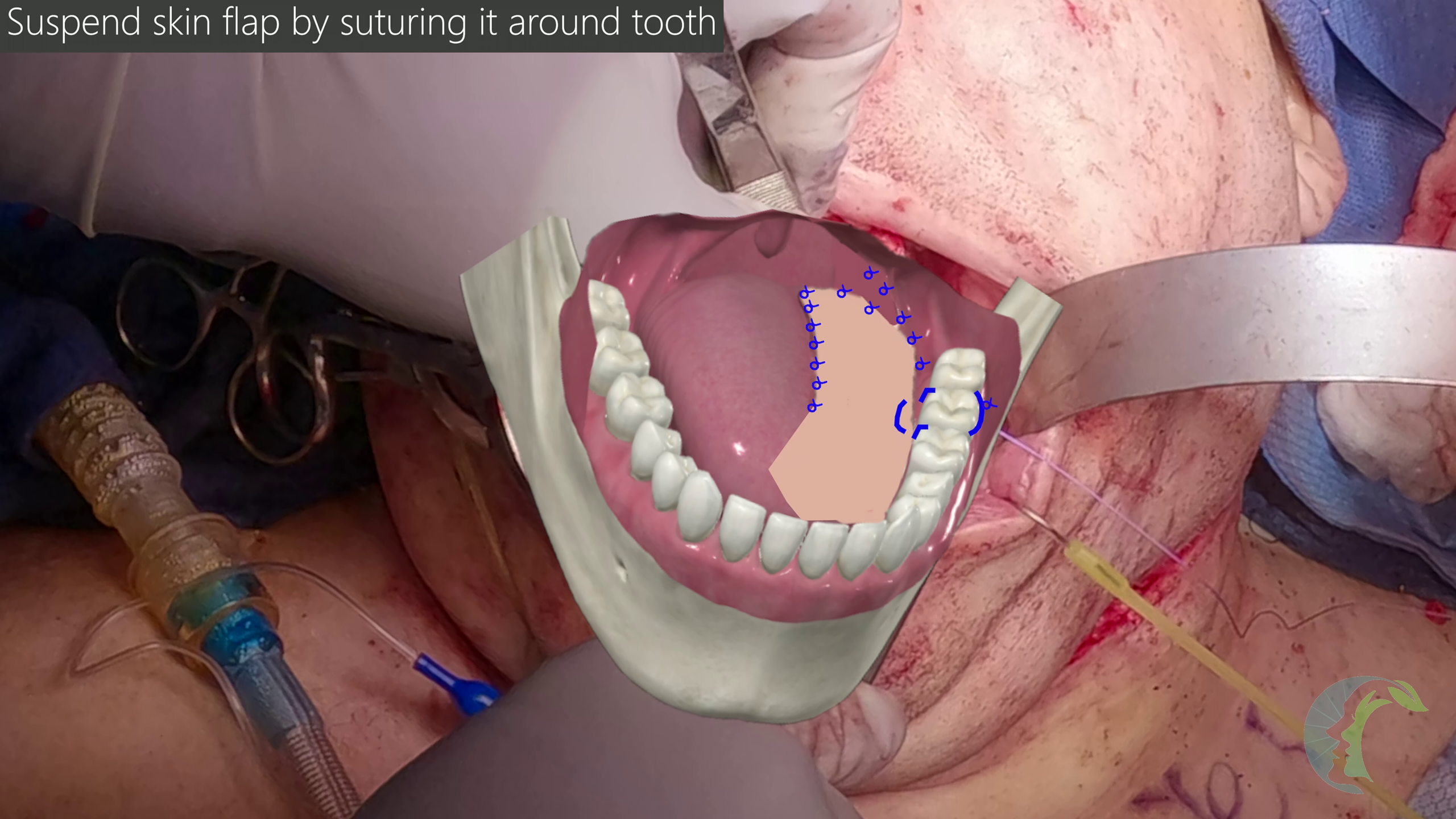
Dr. Gordin prefers to use PDS sutures for circumdental sutures as it offers greater elasticity and may provide greater watertight closure as tissues swell [5].
5.5 Final Checks
Ensure a watertight seal at the flap–tongue–floor-of-mouth junctions and adjacent to remaining native mandible or teeth as these locations are prone to salivary leaks.
Once vessel anastomosis is complete, confirm adequate Doppler signals. Drs. Lee and Kandl prefer to use vein doppler couplers (Flow coupler system, Synovis MCA).
If significant flap swelling is present, lace a bite block for approximately 5 days to prevent dental trauma to native tongue or flap [1]. Dr. Gordin prefers to use a soft bite block with rolled up moistened 4x4s [5].
6 Surgeon-Specific Pearls
In this section, Dr. Gordin and Dr. Kandl share their insights and their surgical pearls [5,6].
Dr. Chris Kandl:
Dr. Eli Gordin: