Segmental Mandibulectomy and Non-Vascularized Tissue-Engineered Reconstruction
Yotom Rabinowitz, DDS, Thomas S. Lee, MD
Published 9/04/2026
Surgical Indications and Patient Selection
-
Non-vascularized tissue-engineered reconstruction is strictly limited to non-radiated candidates who present with intact or minimal intraoral mucosa or external skin defects, allowing for tension-free, watertight primary closure.
- Patients requiring postoperative radiation therapy should undergo free flap reconstruction instead of non-vascularized grafting.
-
Patients presenting with a history of prior head and neck radiation, an anticipated need for post-operative radiotherapy, or extensive soft tissue defects are excluded from this technique and require microvascular free tissue transfer, such as a fibula free flap.
Relevant Surgical Anatomy and Fascial Layers
-
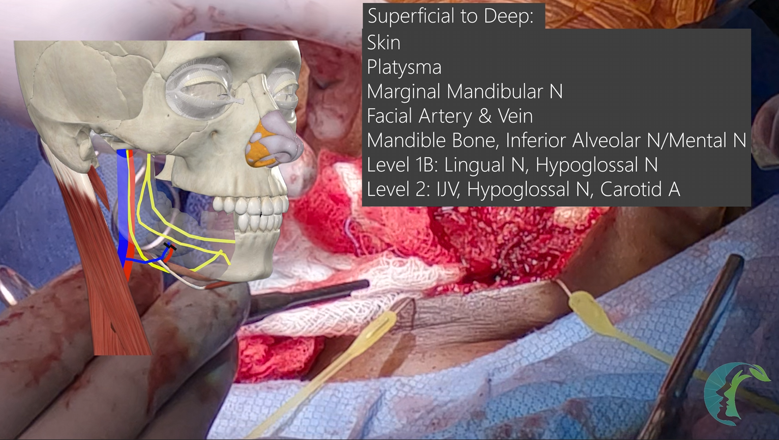
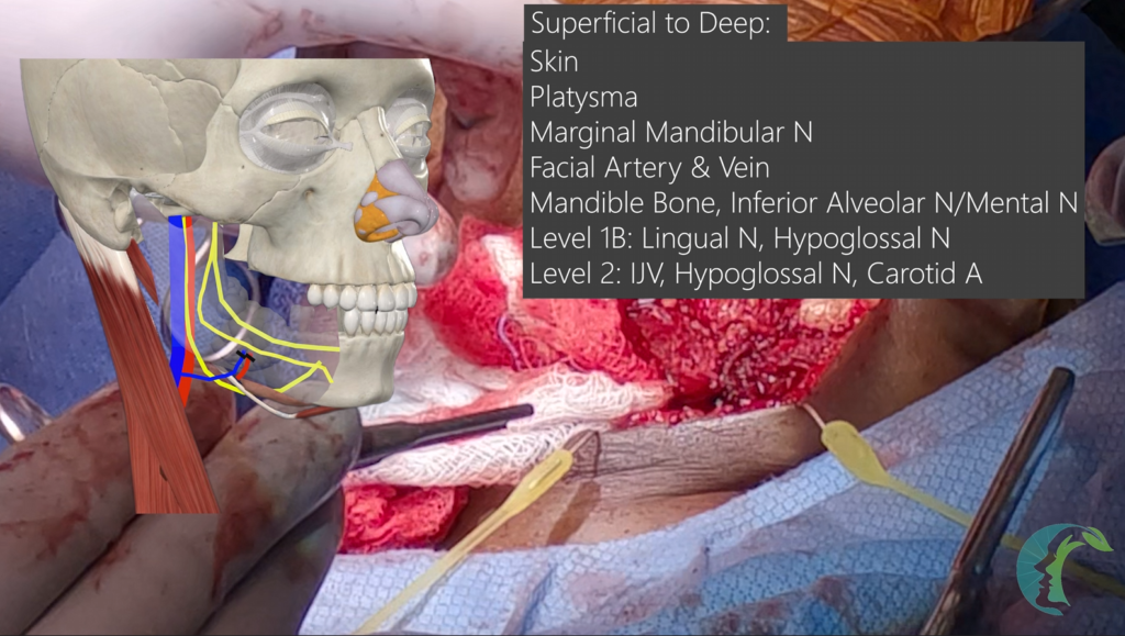
Superficial Layers and Fascia: Moving from superficial to deep, the anatomy comprises the skin, subcutaneous fat, and the platysma muscle.
-
Subplatysmal Tissue Plane (deep to platysma): The marginal mandibular nerve (a branch of the facial nerve) runs deep to the platysma muscle and sits directly superficial to the submandibular gland capsule. This nerve should be identified, and in this case, it will be raised with the skin flap over the mandible to preserve it. The facial artery and vein course inferiorly to the mandible and continues superior to the mandible to become the angular artery. The marginal mandibular nerve will be located superficial to the facial vein at varying locations. The general rule of cutting the facial vein two fingerbreadths below the mandible to protect the nerve is very crude and not accurate, as the point where the marginal mandibular nerve crosses the vessels varies greatly.
Mandible: Within the bone, the inferior alveolar nerve enters the proximal mandible and exits the bone at the mental foramen as the mental nerve. The inferior alveolar nerve and lingual nerve arise from CN V3.
Deep to Mandible: Level 1B and Level II lymph node zones are situated deep to the mandible.
-
Level 1B houses the submandibular gland, the submandibular lymph nodes, and the lingual nerve as it enters the floor of the mouth. The hypoglossal nerve, located deep to the digastric muscle, also arises from Level II and travels along the Level 1B region inferior to the lingual nerve. You will typically encounter the lingual nerve along the floor of the mouth, and it is located within the submucosal tissue plane and is prone to injury if caution is not exercised.
Level 2: The hypoglossal nerve provides motor innervation to the tongue and is identified within Level II/1B zones deep to the internal jugular vein and superficial to the carotid artery structures. Within Level II, the internal jugular vein is located superficially.Deep to the internal jugular vein (IJV) lies the hypoglossal nerve.Deep to the hypoglossal nerve lies the carotid artery.
Patient History:
-
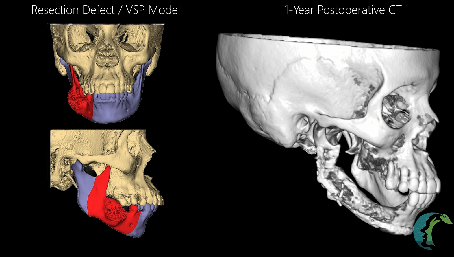
Patient Presentation: 52-year-old female presenting with a mandible lesion biopsied as low-to-intermediate-grade mucoepidermoid carcinoma.
-
Surgical Plan: Segmental mandibulectomy with mucosal margins and sacrifice/reconstruction of the inferior alveolar nerve.
-
Contingency Planning: Free flap reconstruction was prepared in case of inadequate mucosa for tension-free closure.
-
Radiation Considerations: Patients requiring postoperative radiation therapy for high-grade mucoepidermoid carcinoma are planned for a primary fibula free flap instead.
Bone Marrow Aspirate Concentrate (BMAC) Harvest
-
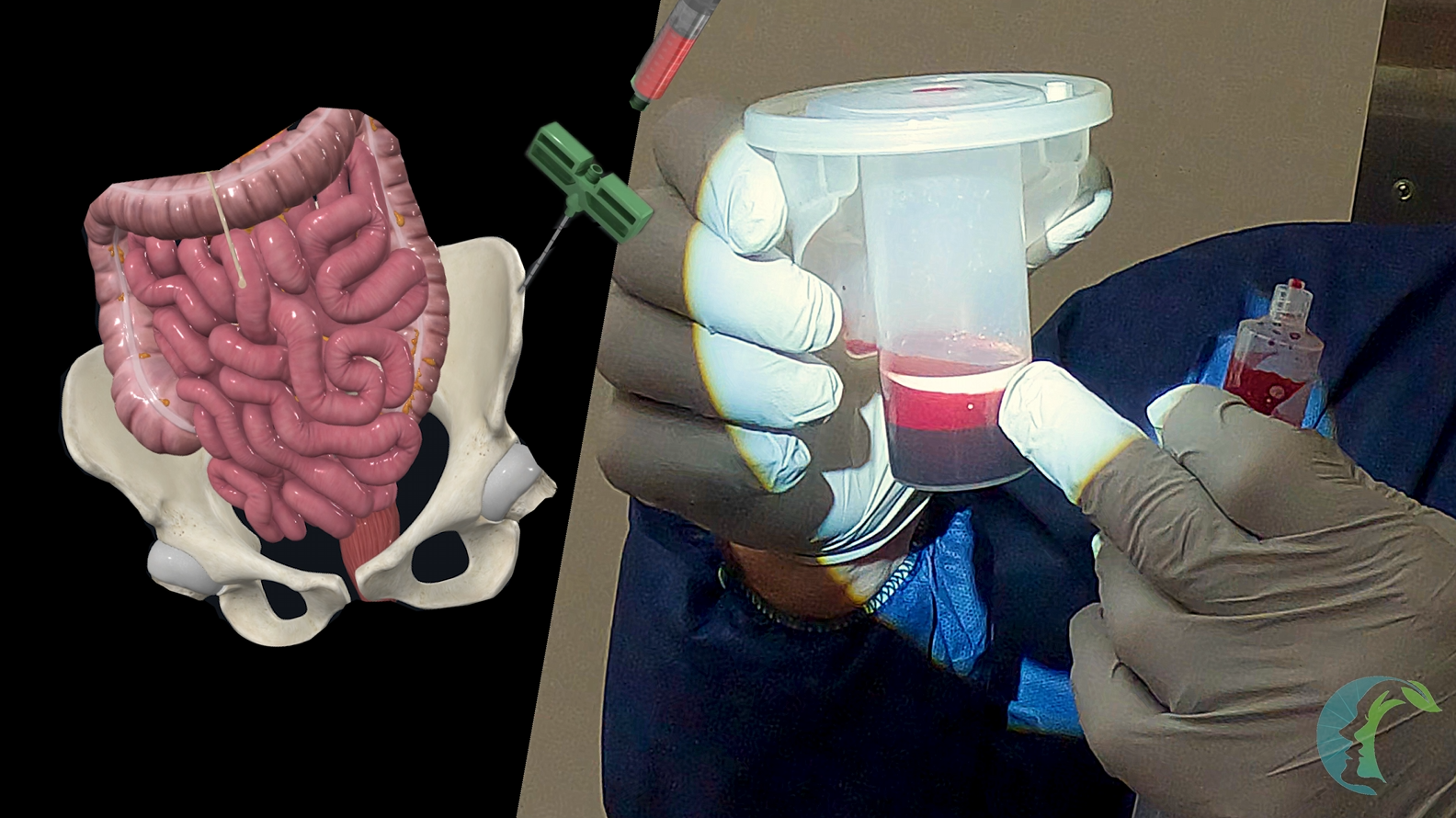
Aspiration Approach: The anterior iliac crest is palpated, and a long needle is introduced into the marrow space using a mallet, maintaining a trajectory strictly between the inner and outer cortices to avoid the inner cortex and prevent bowel injury.
-
Optimization of Cellular Yield: A syringe is attached to create a negative pressure vacuum; if initial aspirate yield is poor, the needle tip is repositioned slightly shallower (withdrawn by a few centimeters) or redirected posteriorly rather than anteriorly to capture a high concentration of mesenchymal stem and progenitor cells.
-
Processing: The harvested marrow aspirate is spun down in a fresh centrifuge to separate its distinct biological layers before being combined with cellular allograft matrix.
Intraoral Prepping:
-
Intermaxillary Fixation (IMF) Placement: Intermaxillary fixation (IMF) screws are placed in the maxilla and mandible away from the operative site. Maxillomandibular fixation will be performed after the tumor resection.
-
Mucosal Incision: A mucosal incision is performed. In this case, since we are dealing with a malignant condition (mucoepidermoid carcinoma), around 1 cm of mucosal margin is achieved. The mucosal incision is made along the medial and lateral aspects of the abnormal mucosa.
-
Sterile Draping: The patient’s mouth is then sealed off using Ioban. The patient is prepped and draped sterilely with povidone-iodine.
Cervical Approach and Soft Tissue Dissection
-
Soft tissue margin: Soft tissue around the tumor is kept down on the tumor for soft tissue margins. Unlike in a benign lesion where a subperiosteal tissue plane can be maintained, as much soft tissue as possible is left on top of the tumor. The marginal mandibular nerve is lifted while leaving the rest of the soft tissue on top of the tumor for soft tissue margins. Dissection is continued from the inferior to the superior border of the tumor until the previously made intraoral mucosal incision is encountered.
Mandibular Osteotomies and Neural Management
-
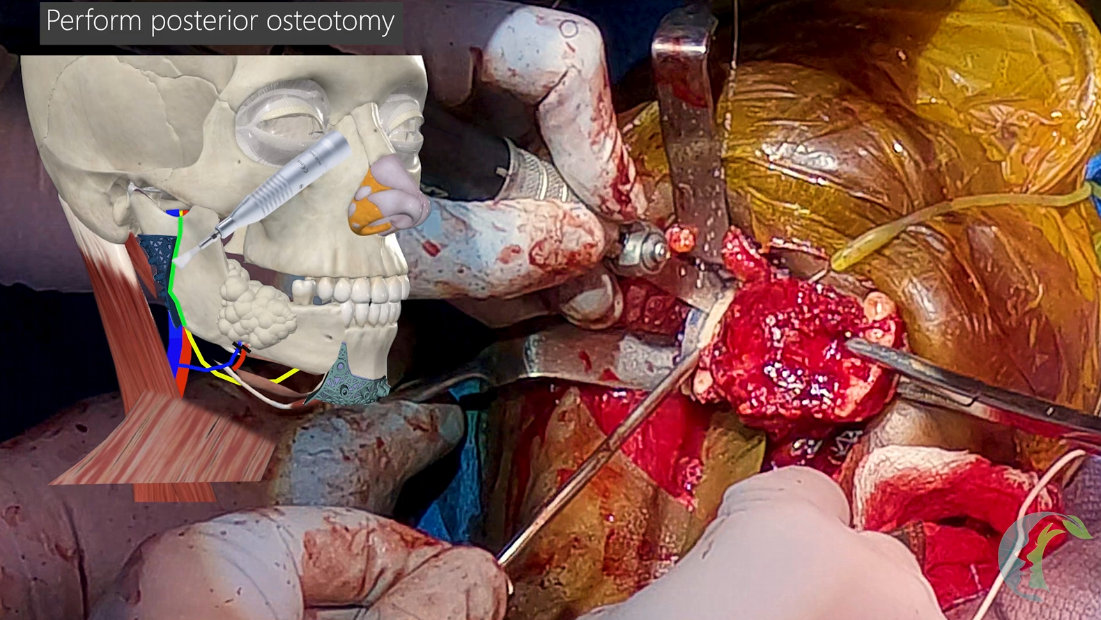
Cutting Guides: Custom titanium cutting guides matching the inferior and posterior borders of the mandible are secured via monocortical screws, followed by bicortical predictive drilling. If exposure of the posterior mandible is difficult for the posterior cutting guide placement, one can consider making the anterior mandibular osteotomy first to advance the mandible forward for greater exposure.
-
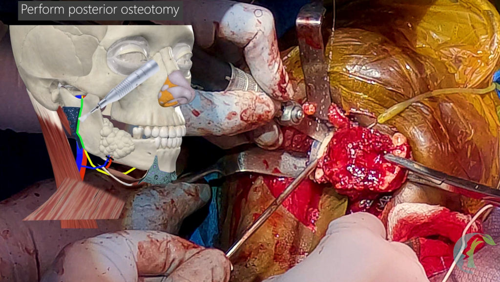
Osteotomy Execution: The anterior mandibulectomy is performed first to allow anterior mobilization of the condyle segment. The posterior segment exposure involves releasing soft tissue attachments to the sigmoid notch, removing the coronoid process entirely to eliminate temporalis muscle tension/pull on future hardware, and executing the osteotomy with a side-cutting bur and T-bar osteotome.
-
Nerve Reconstruction: The proximal inferior alveolar nerve and distal mental nerve ends are identified and tagged with Prolene sutures. Frozen sections are sent to confirm negative perineural invasion.
Watertight Mucosal Closure: To eliminate salivary contamination and prevent graft infection, a meticulous multi-layered closure of the oral mucosa is performed starting at the retromolar trigone.
-
Sharp bone edges along the superior margin of the mandible are drilled down with a round cutting bur to eliminate any sharp points that could cause inadvertent mucosal perforation, dehiscence, or salivary leakage.
-
Using a 3-0 PDS suture in a running, inverted fashion, the native floor of the mouth is re-established from a posterior to anterior direction to ensure a robust, tension-free watertight barrier between the oral cavity and the neck. This will be the first layer of mucosal closure. Mucosal edges should be inverted into the intraoral cavity.
-
The second layer consists of layering submucosal soft tissue that is advanced to reinforce the closure. Masseteric muscle or buccal fat along with other soft tissue can be incorporated deep to the first layer closure. Care must be taken to avoid catching the lingual nerve, remaining inferior alveolar nerve, and the marginal mandibular nerve.
-
You irrigate the neck with povidone-iodine and close the neck loosely with staples and seal it off with Ioban.
-
We break through previously placed Ioban over the mouth and enter the intraoral cavity.
-
The third layer, consisting of the intraoral mucosal layer, will be performed once we enter the mouth.
-
Patient is placed into maxillomandibular fixation to achieve premorbid occlusion. A custom splint can also be used to achieve new occlusion if desired.
-
Additional IMF screws are placed centrally if the tumor-side has occlusion that opens.
- Patient is once again prepped with povidone-iodine and the mouth is sealed off. Neck ioban seal is broken and begin reconstruction.
Inferior Alveolar Nerve Repair Using Nerve Tape: Restoring the inferior alveolar nerve should restore lower lip sensation.
- The proximal inferior alveolar nerve and distal mental nerve ends are trimmed back to fresh bleeding margins (~1–2 mm trimmed back) and prepared for reconstruction. Using a sutureless nerve connector (Nerve Tape, Biocircuit Technologies, Atlanta, Georgia, USA ), the allograft cable graft (Avance, Axogen, Alachua, Florida, USA) is preloaded. The proximal inferior alveolar nerve stump is secured to bridge the defect without tension. The entire construct is then swept toward the distal mental nerve, where a 1-millimeter gap is maintained to allow axons to line up before the nerve tape is wrapped securely around the coaptation site to encourage directional axonal regeneration.
- If nerve tape is not available, nerve coaptation can be performed by lining up the epineurium with 8-0 nylon sutures.
- If an allograft cable graft is not available, one can consider using the great auricular nerve or sural nerve as the donor nerve.
Non-Vascularized Bone Graft Insertion with PRP and Fibrin:
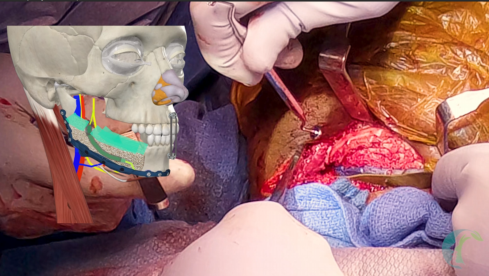
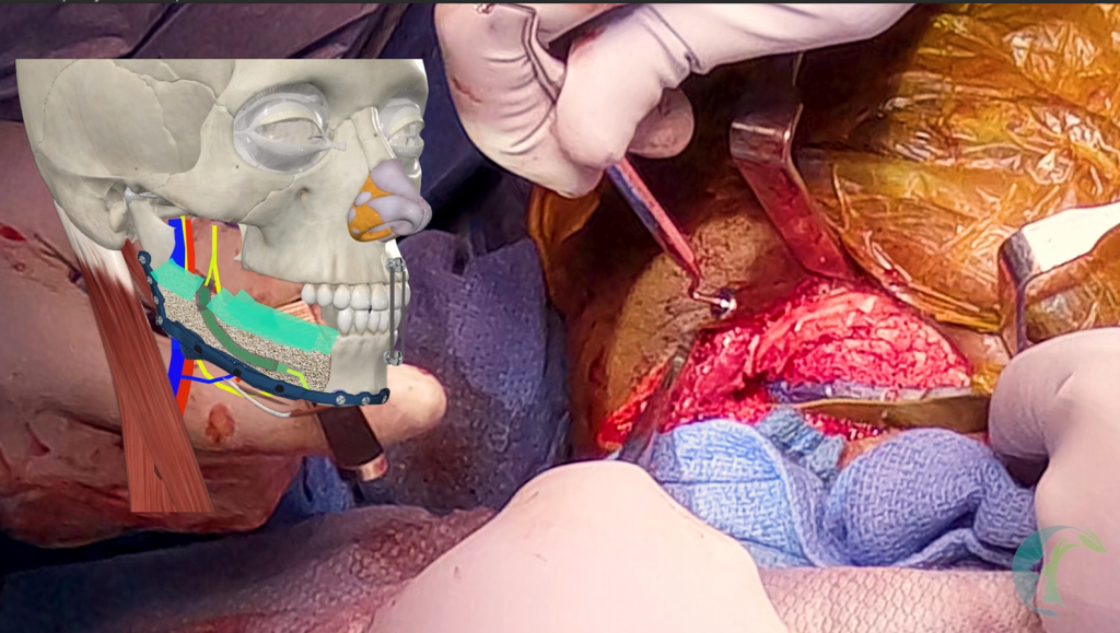
- The custom titanium crib plate is secured to the remaining condyle and parasymphysis using bicortical non-locking screws while the patient is locked into pre-morbid occlusion via IMF wires.
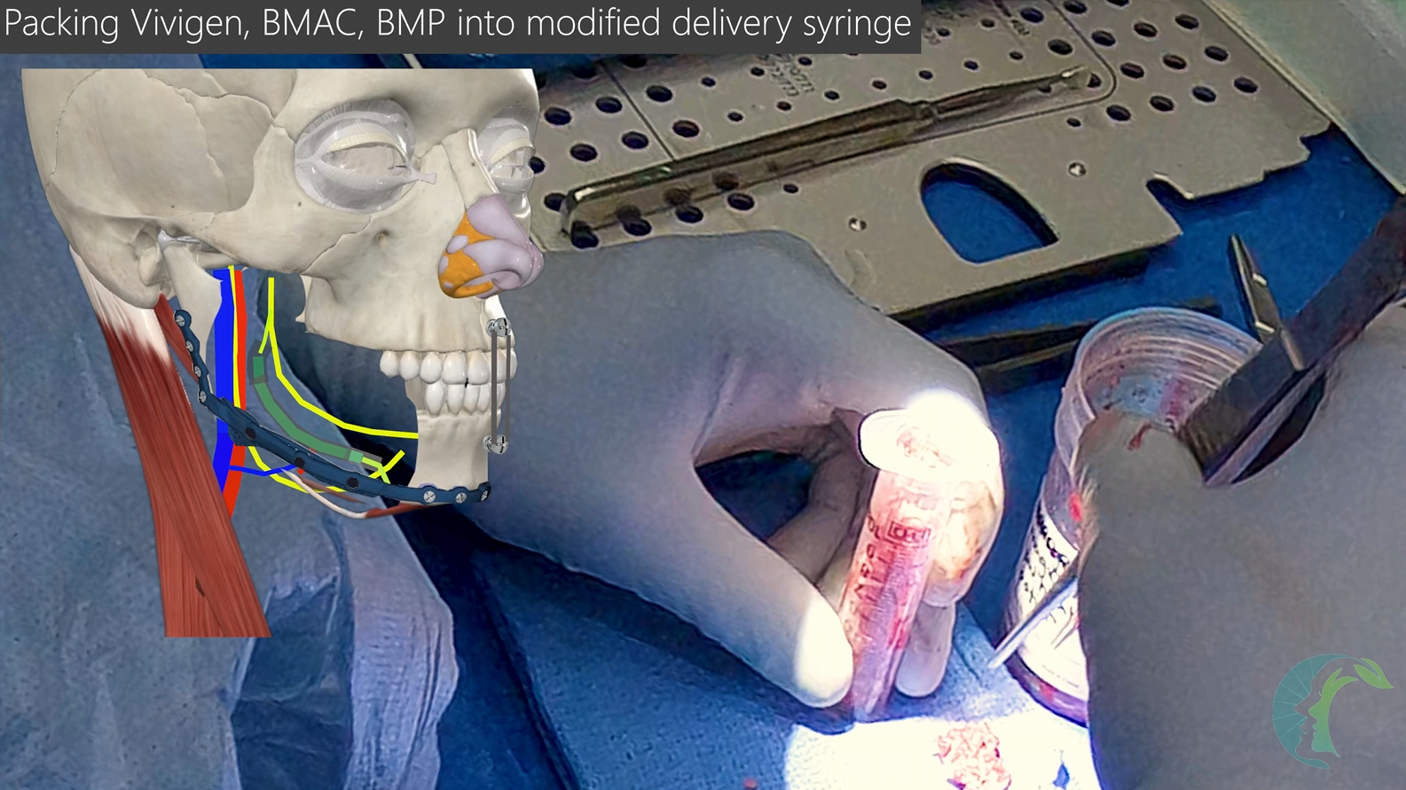
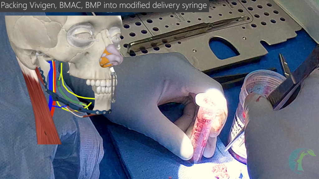
- A composite tissue-engineered matrix consisting of Vivigen cellular bone allograft, recombinant human bone morphogenetic protein (BMP), bone marrow aspirate concentrate (BMAC), and platelet-rich plasma (PRP) is serially packed from inferior to superior into the crib plate, positioning the nerve graft buccally or inferiorly to avoid interference with future dental implant placement.
- PRP is compressed into a jelly consistency and laid deep to the mucosal closure to provide an additional layer against leakage.
- Fibrin glue is sprayed from top to bottom over the nerve repair and bone graft complex to consolidate and immobilize the materials.
Post-Operative Protocol:
- Surgical drains are intentionally omitted to prevent the vacuum system from aspirating growth factors, BMAC, or graft material from the construct.
- The skin flap is closed in layers over the immobilized site.
- Patient is maintained in maxillomandibular fixation on a liquid diet for a minimum of 2 to 4 weeks to promote complete graft stabilization and early revascularization. Maintaining rigid MMF is crucial while the bone graft consolidates.
- At the 3- to 4-week mark, provided healing is uneventful, all wires and IMF screws are removed.