MASTERCLASS IN TOTAL TMJ RECONSTRUCTION (TMJR)
Faculty: Louis G. Mercuri, DDS, MS, FRCSDS (Eng) (Hon)
Adjunct Professor
Department of Orthopaedic Surgery, Rush University Medical Center, Chicago, IL
SECTION 1: PREOPERATIVE EVALUATION, PATIENT SELECTION, AND SYSTEMIC CONSIDERATIONS
1.1 Biopsychosocial Overlay & Chronic Pain Dynamics
Patients presenting for temporomandibular joint (TMJ) total joint replacement frequently present with significant biopsychosocial overlays. A history of multiple prior interventions may be common. Dr. Mercuri’s patients had on average 5.4 surgeries prior to total joint consultation with him (Mercuri et al, 1995). In patients with multiple surgeries and chronic surgical trauma, central pain sensitization may occur, shifting nociception from peripheral articular structures to central mechanisms and making perioperative pain control difficult to manage (La Touche et al., 2018).
Surgical Outcomes Expectation Setting
The primary goals of surgical intervention are anatomical restoration, airway stabilization, and functional improvement (e.g., mastication and speech). Pain reduction should be discussed using realistic expectations based on the patient’s surgical history. Dr. Mercuri’s clinical experience suggests the following approximate expectations for pain reduction; these figures should not be interpreted as validated predictive estimates:
- No Prior Surgery Cases: Up to 75% pain reduction anticipated.
- 3–5 Prior Surgeries: Approximately 50% pain reduction.
- 5 Prior Surgeries: Maximum expected pain reduction drops to ~25%.
Pain relief must never be guaranteed. The primary goal of TMJR is restoration of mandibular function; any pain relief should be considered a secondary benefit, particularly in the multiply operated patient.
1.2 Indications and Patient Selection
Optimal Candidates
Indications for alloplastic temporomandibular joint replacement are informed by established clinical criteria and published literature, including the following (Mercuri, 2016; Mercuri & Granquist, 2022; Mercuri, 2023; Mercuri, 2028):
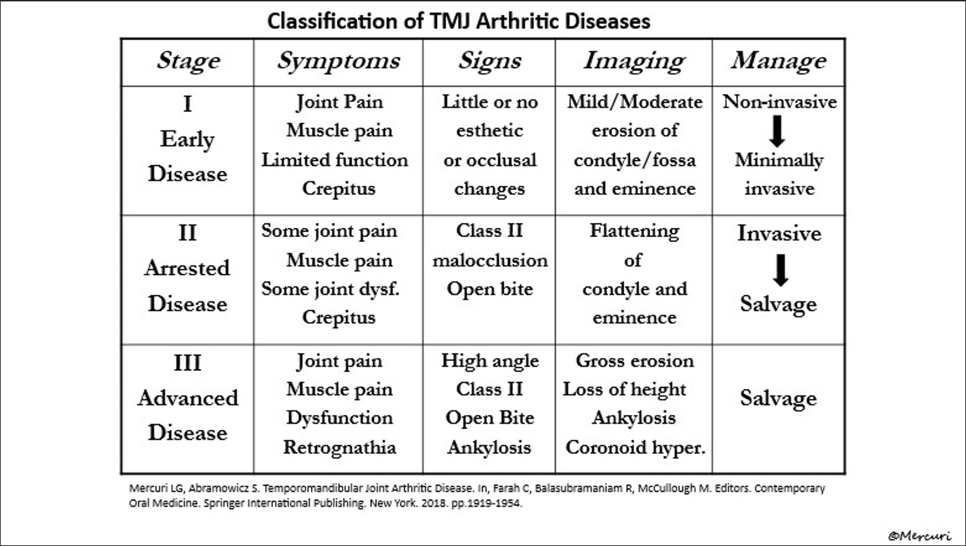
- Advanced Stage Osteoarthritis (Figure 1; Mercuri & Abramowicz, 2018).
- End-stage rheumatoid arthritis or inflammatory arthritis. Dr. Mercuri considers these patients appropriate candidates when disease status, systemic health, and treatment expectations are favorable.
- Ankylosis (bony or fibrous).
- Avascular necrosis / End-stage condylar resorption.
- Failed autogenous or alloplastic joint reconstructions.
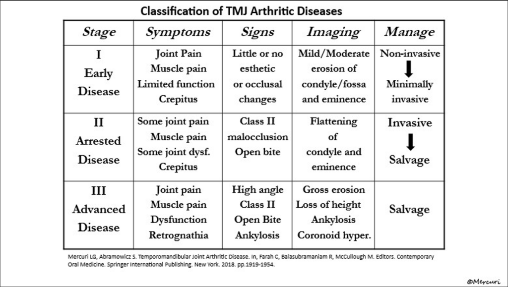
Figure. Classification of TMJ Arthritic Diseases (Adapted from Mercuri & Abramowicz, 2018).
Relative Indications & Pediatric/Skeletally Immature Considerations
- Skeletally Immature Patients: In patients who have reached skeletal maturity—often approximately 15 years or older in females and 17 years or older in males—standard patient-fitted TMJR protocols may be considered. Individual skeletal maturity should be assessed rather than relying solely on chronological age. In skeletally immature patients (e.g., those with post-traumatic ankylosis), the primary goal is establishing continuous postoperative joint mobility. This approach is consistent with Moss’s Functional Matrix Theory, which emphasizes the important role of the functional demands and mechanical environment of the surrounding soft tissues in regulating skeletal growth and remodeling. This rationale is consistent with aspects of Moss’s Functional Matrix Theory, which proposes that functional demands and the mechanical environment of surrounding soft tissues influence skeletal growth and remodeling. In skeletally immature patients, restoration of mandibular function may therefore provide a more physiologic mechanical environment for continued growth and remodeling (Moss, 1997).
- Connective Tissue Disorders: Conditions such as Ehlers-Danlos Syndrome can undergo TMJR, though soft tissue stability must be evaluated.
- Diabetes Mellitus: Controlled diabetes is acceptable with optimized HbA1c.
Contraindications
- Post-Radiation Osteoradionecrosis & Extended Defects: Patients with prior head/neck radiation combined with extensive oncologic resections/defects are not ideal candidates for alloplastic TMJR. Vascular compromise and compromised mucosal integrity can lead to high rates of wound breakdown, hardware exposure, and periprosthetic joint infection (PJI). In these scenarios, vascularized tissue reconstruction may be favored over alloplastic TMJR depending on the extent of the defect, radiation injury, soft-tissue condition, and reconstructive goals. Microvascular free tissue transfer, such as an osteocutaneous fibula free flap, is one reconstructive option.
- Active Infection: Local or systemic active infection.
1.3 Dental Optimization and Extraction Protocol
- Severe Dental Disease: Dr. Mercuri recommends postponing TMJR for approximately two to four weeks after dental extractions when feasible, allowing adequate mucosal healing before joint replacement. Timing should be individualized according to the extent of dental disease and healing.
- Ankylosis Patients: Patients with ankylosis frequently present with poor oral hygiene and severely decayed or impacted teeth due to their inability to open their mouths, creating a nidus of infection. For these patients, a staged intraoperative approach is required. The surgeon must first release the ankylosis to achieve mouth opening. The oral cavity is then prepped with povidone-iodine, the necessary extractions are performed, and the sockets are closed. Following the intraoral stage, the patient must be completely re-prepped and re-draped before proceeding with the TMJR hardware implantation.
SECTION 2: INFECTION CONTROL PROTOCOLS & MATERIAL SCIENCE
2.1 Periprosthetic Joint Infection (PJI) Pathophysiology
PJI occurs in approximately 3% to 5% of TMJ replacement cases, consistent with infection rates reported in several TMJR series (e.g., 4.5% in a 10-year retrospective analysis of 178 TMJ prostheses) (McKenzie & Louis, 2017). The primary causative pathogens are Staphylococcus aureus, Staphylococcus epidermidis, and Cutibacterium acnes.
Cutibacterium acnes Nuances
- Localization: Unlike Staphylococcus species, C. acnes resides deep within sebaceous glands and hair follicles. Topical cutaneous antiseptics cannot fully eradicate follicular bacterial reservoirs. Incisions through hair-bearing skin transsect these follicles, releasing bacteria into the deep surgical field
- Hydrogen peroxide may provide additional reduction of Cutibacterium acnes burden when incorporated into standard surgical skin preparation. In a prospective controlled study of shoulder arthroplasty, Chalmers et al. found that adding hydrogen peroxide to standard skin preparation reduced the proportion of patients with multiple positive intraoperative cultures and reduced the frequency of positive glenohumeral joint cultures, without observed adverse skin reactions. Although these findings support hydrogen peroxide as an adjunct for reducing acnes contamination, direct evidence that this approach reduces PJI in TMJR is lacking. Accordingly, its use in TMJR should be considered an extrapolation from prosthetic shoulder surgery rather than an established TMJR-specific infection-prevention strategy (Chalmers et al., 2019).
- Microbiology Protocol: Because C. acnes is slow-growing, prolonged anaerobic incubation—often at least 14 days—may improve detection when PJI is suspected. The microbiology laboratory should be notified when prolonged incubation is desired.
- Biofilm Mechanics: Biofilms form on both Ultra-High-Molecular-Weight Polyethylene (UHMWPE) and metallic surfaces (Mercuri, 2006). Established biofilm formation makes successful debridement-and-implant-retention strategies less reliable, and management often requires complete device removal with staged or immediate replacement depending on the clinical circumstances. Dr. Mercuri recommends complete device removal when an established prosthetic biofilm is suspected rather than attempting debridement and implant retention.
2.2 Antimicrobial Prophylaxis & Prep Protocol
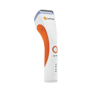
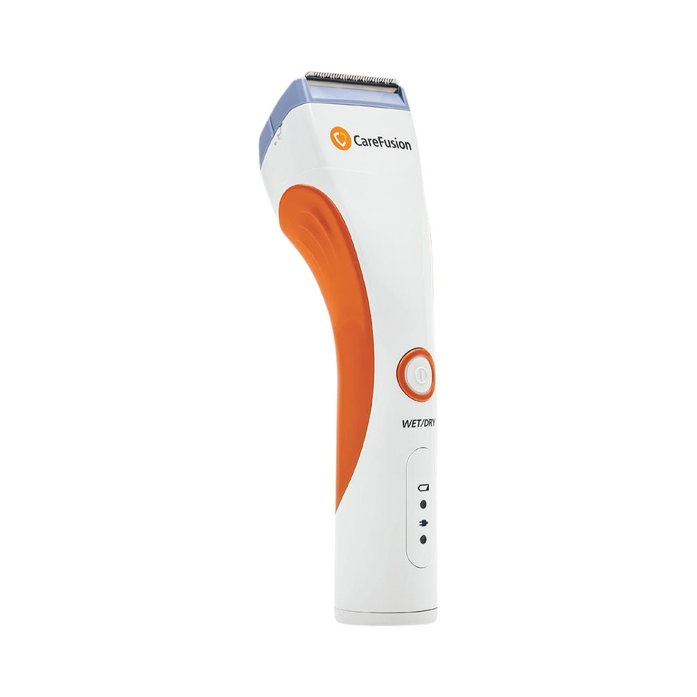
- Pre-op Hair Prep: Electric clippers ONLY. Avoid razors; manual shaving creates epidermal micro-lacerations that serve as entry points for S. aureus and S. epidermidis.
- Systemic Prophylaxis: Dr. Mercuri’s antimicrobial protocol includes preoperative IV cefazolin, with alternative agents (IV Clindamycin) selected for patients with relevant antibiotic allergies. Antibiotics are administered before incision and redosed intraoperatively according to standard pharmacokinetic principles. His protocol also includes postoperative oral antibiotics for approximately 7–10 days; postoperative antibiotic duration should be individualized according to institutional protocols and patient-specific factors.
- Skin Preparation: Povidone-Iodine solution. Povidone-iodine has demonstrated efficacy as an intraoperative antiseptic in orthopedic total joint arthroplasty and is incorporated into Dr. Mercuri’s TMJR infection-control protocol.
- Component Soaking & Antiseptic Irrigation (Machinski et al. Meta-Analysis): A meta-analysis by Machinski et al. evaluated antiseptic irrigation consisting of chlorhexidine or povidone-iodine compared to normal saline after primary total joint arthroplasty in orthopedics (Machinski et al., 2025). Reviewing 11 studies comprising 67,742 patients, the baseline infection rate was 1.02%. Among the 24,025 patients who received antiseptic irrigation (chlorhexidine or povidone-iodine), the infection rate dropped significantly to 0.86%. Both povidone-iodine and chlorhexidine demonstrated a risk ratio of 0.6 compared to saline, with no statistical difference between the two solutions. Clinical takeaway: In primary hip and knee arthroplasty, intraoperative irrigation with povidone-iodine or chlorhexidine was associated with a lower risk of PJI compared with saline irrigation. Whether these findings translate directly to TMJR requires clinical extrapolation.
- Vancomycin Powder: Dr. Mercuri incorporates intrawound vancomycin powder into his infection-control protocol, applying it to the deep wound cavity before closure to provide local coverage against gram-positive organisms, including C. acnes.
2.3 Material Science & Biomechanics
Fossa Component
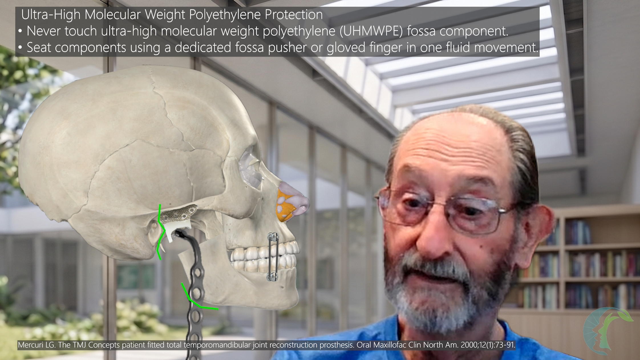
- Composed of an Ultra-High-Molecular-Weight Polyethylene (UHMWPE) articular surface bonded to a commercially pure titanium mesh backing.
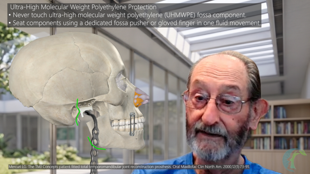
- Polyethylene Care: Avoid contacting or scuffing the UHMWPE articular surface with metallic instruments; Dr. Mercuri recommends using dedicated plastic pushers or digital pressure when seating the component. Micro-scratches accelerate abrasive wear, producing polyethylene debris that may trigger macrophage-mediated osteolysis. Use dedicated plastic fossa pushers or digital pressure to seat the component.
Ramus Component
- Composed of a Cobalt-Chromium-Molybdenum (CoCrMo) alloy condylar head mated to a Titanium Alloy (Ti6Al4V) ramal body.
- Metal Surface Integrity: Scratching or scuffing the CoCrMo condylar head with retractors or elevators creates surface asperities. These act as sites for potential corrosion and as a rasp against the UHMWPE fossa component, leading to premature wear.
Manufacturing Process Comparison
- The structural integrity and long-term durability of the hardware may differ depending on the fabrication method used to create the prosthesis (Neto et al., 2022).
- Forged / CNC Milled Hardware: Possesses uniform, homogeneous metallurgical grain structures with minimal micro-voids, providing high fatigue strength under cyclic masticatory loading.
- 3D-Printed / Additive Manufactured Metals: Additive manufacturing can produce different microstructures and may introduce variations in porosity and other material characteristics. These differences may influence mechanical and electrochemical behavior and should be considered when evaluating long-term implant performance (Neto et al., 2022).
SECTION 3: ANESTHETIC MANAGEMENT, SURGICAL SETUP, AND AIRWAY CONTROL
3.1 Patient Positioning and Table Configuration (Mercuri, 2011)
- 180-Degree Table Rotation: The operating table is rotated 180 degrees away from the anesthesia machine, positioning the surgeon directly at the head of the bed looking inferiorly over the patient’s face.
- Anesthesia Placement: The anesthesia provider and circuit are located at the foot of the bed.
3.2 Nasal Endotracheal Intubation Protocol (Mercuri, 2011)
- Tube Type: Standard endotracheal tube (NOT a curved nasal endotracheal tube) to allow flexible routing toward the foot of the bed.
- Fixation: Secure the tube to the nasal septum using a 3-0 silk suture, followed by adhesive taping.
- Tissue Protection: Apply Dexamethasone cream liberally around the alar rim and nares to prevent pressure ischemia, friction abrasions, and necrosis during table rotation and surgical manipulation.
- Draping/Isolation: Wrap the breathing circuit connections in sterile gauze/drapes and seal off the oral cavity with a sterile Tegaderm barrier.
3.3 Ocular and Otic Protection Protocols
Eye Care (Mercuri, 2011)
- Irrigate eyes with sterile water.
- Apply ophthalmic ointment.
- Tape eyelids closed.
- Secure rigid plastic eye goggles over the orbits. This prevents accidental elbow or instrument compression on the globes during surgical access.
- Postop Management: Remove goggles, irrigate eyes with sterile water, and instill artificial tears.
Ear Care (Mercuri, 2011)
- Preoperative Otoscopy: Perform otoscopy in the clinic prior to surgery. (Detect foreign bodies, pre-existing tympanic membrane perforations, or cotton pledgets left from prior procedures).
- Intraoperative Packing: Place an antibiotic/steroid-soaked cotton pledget into the external auditory canal (EAC) to block blood and bone debris.
- Postoperative Debridement: Remove the pledget, perform micro-suctioning, and inspect the EAC/tympanic membrane. Ensure there is no disruption of EAC that may have occurred. Disruption of the EAC must be sealed off to prevent hardware infection.
Flush gently with sterile water. Evacuate all blood clots over the tympanic membrane to eliminate postoperative ear pain and vertigo.
SECTION 4: SURGICAL TECHNIQUE & INTRAOPERATIVE PEARLS
Step-by-Step Surgical Workflow
- Preauricular & Transfacial Approaches.
- Fossa Dissection (Dissect to bone medially; stop at medial dip to avoid injuring the middle meningeal artery entering the foramen spinosum).
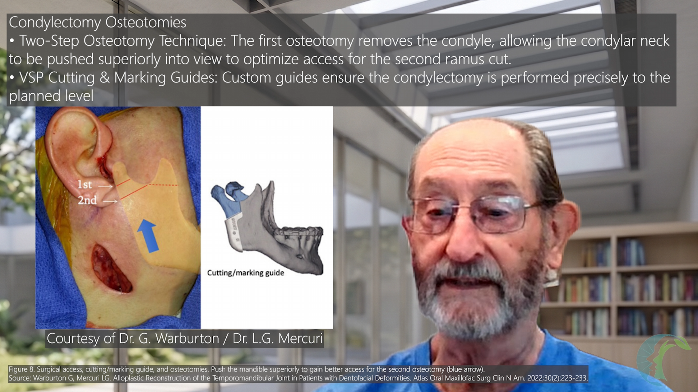
- Condyle Resection (May require two separate osteotomes to allow for the ramus to collapse into the surgical field.).
- Inspect to ensure anterior surface of EAC intact (If disrupted, rotate thin Temporalis Flap. May consider staging the hardware placement after the EAC has fully healed).
- Fossa & Ramus Component Trialing & Seating.
- Low-Speed Irrigation Drilling & Bicortical Screw Fixation.
- Abdominal Fat Graft Harvesting & Packing.
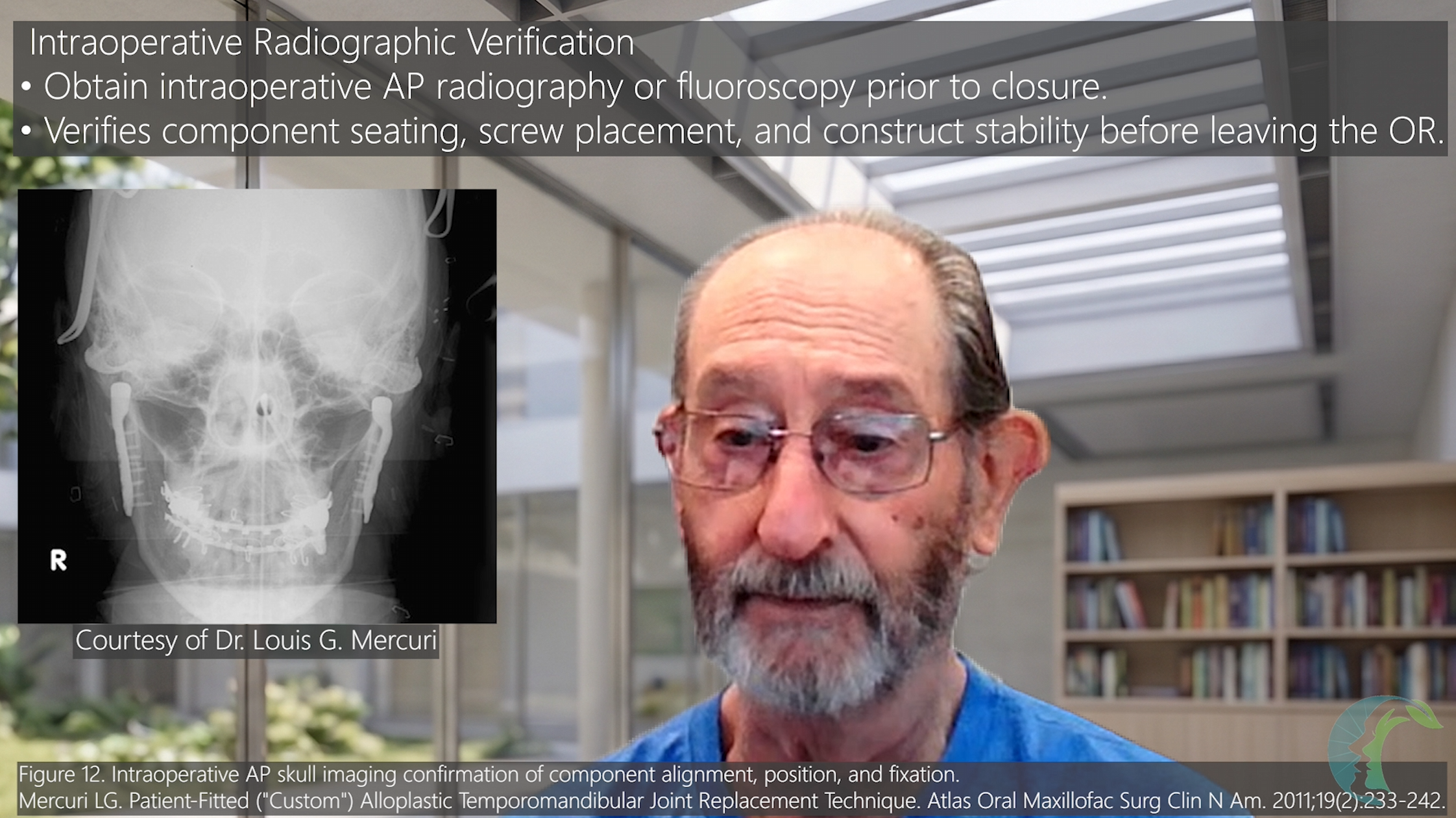
- Fluoroscopy / AP Radiographs to confirm hardware position.
- Hemostasis, Wound Closure & 24-hr Pressure Dressing.
4.1 Fossa Preparation and Anatomic Safety Boundaries
- Complete Soft Tissue Dissection: All disk remnants, fibrous connective tissue, cartilage, and periosteum must be aggressively stripped from the glenoid fossa and articular eminence down to raw bony surface.
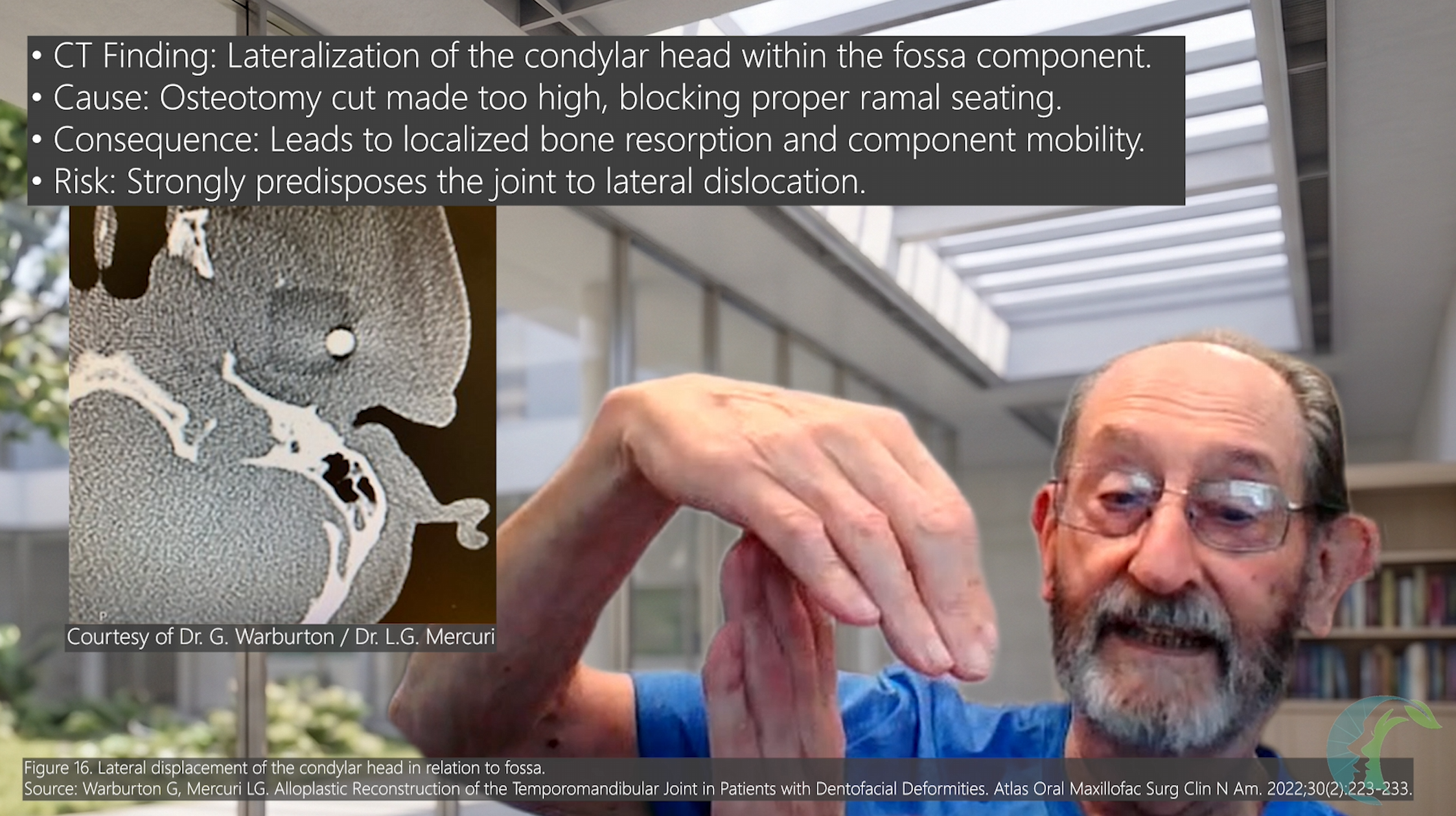
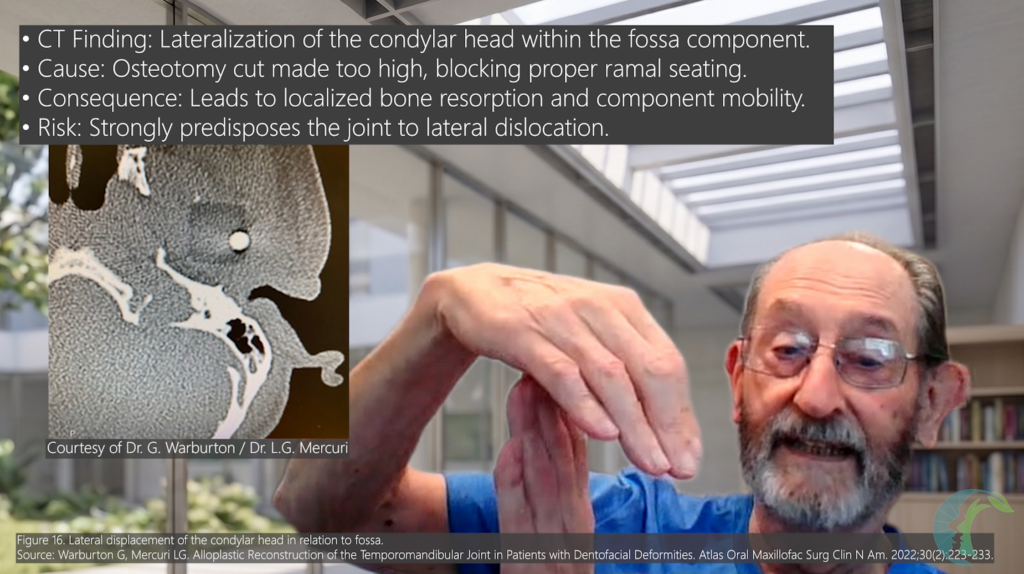
- Critical Pearl: Patient-fitted prostheses are manufactured from 3D bone models reconstructed from CT data (Wolford et al., 2015; Mercuri 2012). CT scans detect only mineralized bone, not soft tissue. Leaving soft tissue in the fossa prevents full seating of the component, forcing it to sit “proud”. This causes condylar head lateralization, non-ideal biomechanics, accelerated UHMWPE wear, and instability.
- Medial Dissection & Foramen Spinosum Margin:
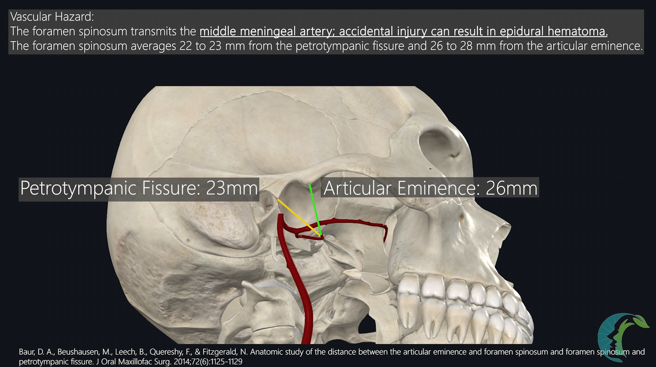
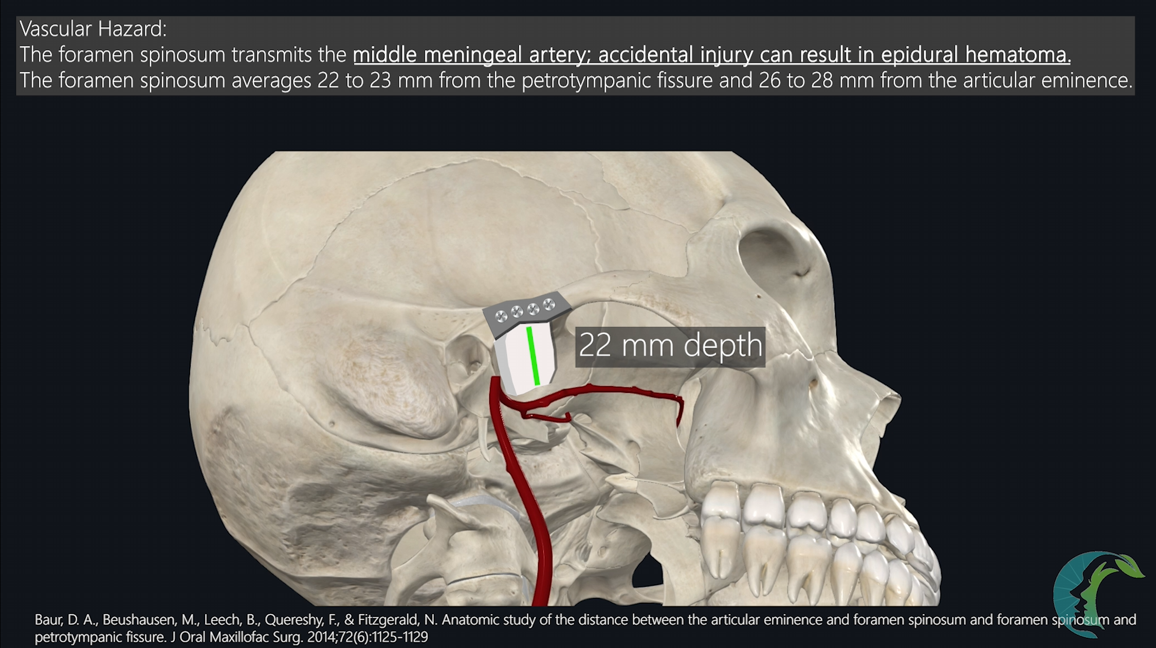
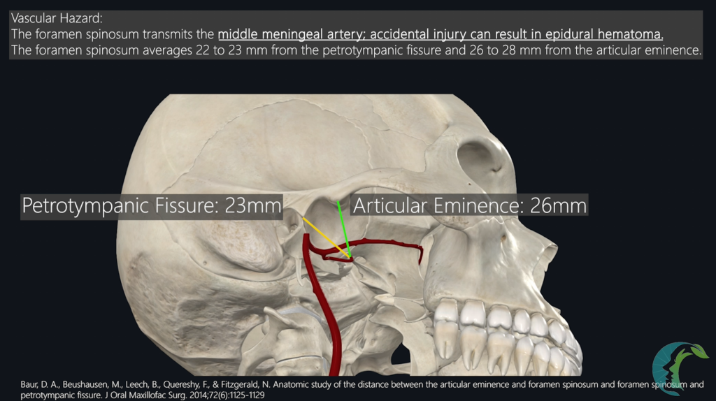
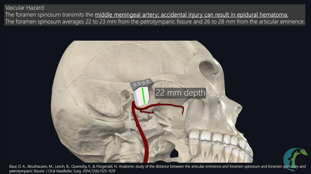
- Foramen spinosum transmits the middle meningeal artery. An injury to the artery can lead to an epidural hematoma, so it should be preserved.
- Dissect medially along the roof of the glenoid fossa.
- The Foramen Spinosum (and the Middle Meningeal Artery) lies approximately 23mm to 25mm medial to the lateral border of the zygomatic arch.
- Custom fossa components typically extend medially to a depth of 22mm, leaving a 1–3mm safety margin.
- Anatomic Landmark: As dissection proceeds medially, the fossa roof exhibits an inferior dip. This dip marks the medial boundary transition toward the foramen spinosum. Cease medial osteoperiosteal stripping at this dip to protect the Middle Meningeal Artery and ensure proper fossa fit.
4.2 Handling External Auditory Canal (EAC) Disruption
- Identification: Direct intraoperative visualization via otoscopy or trans-incisional inspection. Severe ankylosis resection frequently involves anterior tympanic plate fusion and creates the potential for EAC disruption when the ankylosed condyle is drilled away from the tympanic plate.
- Management / Reconstruction:
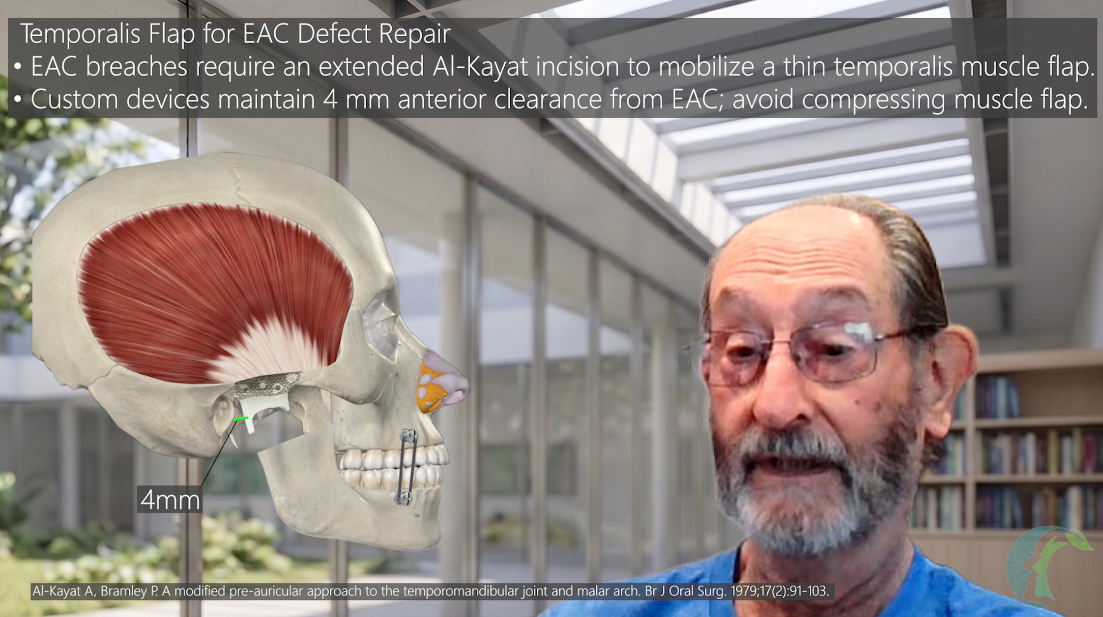
- Custom fossa is designed with a minimum 4mm anterior clearance from the EAC.
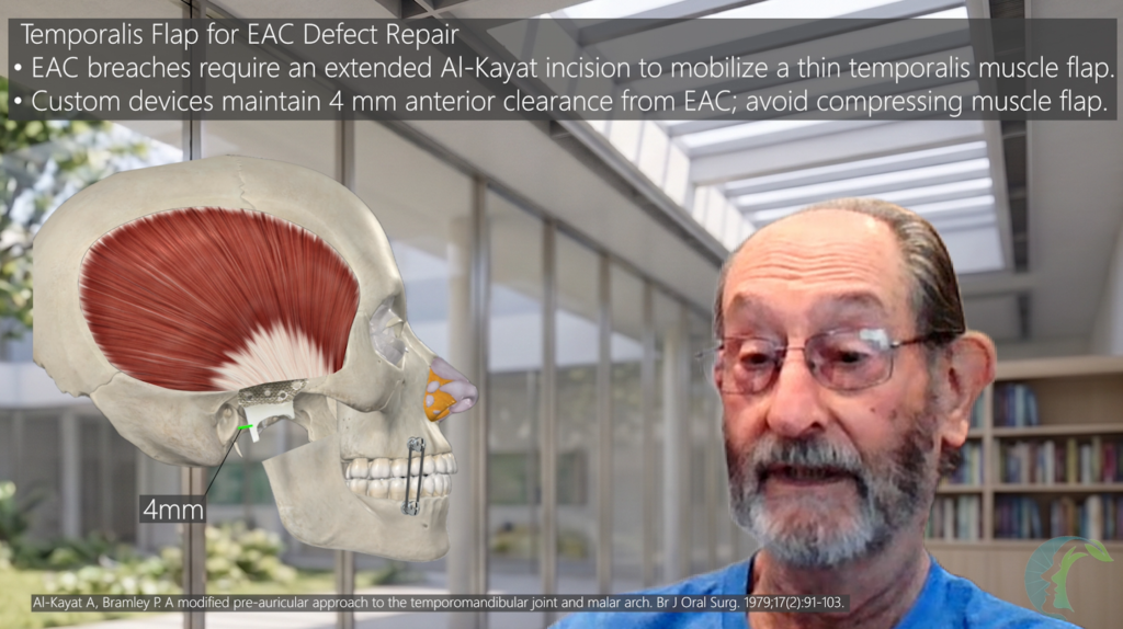
- If the EAC cartilage or skin is breached, elevate a thin, pedicled Temporalis Muscle Flap. EAC must be separated away from the fossa component as the EAC content and skin debris can lead to hardware infection.
- Pivot the superior margin of the temporalis flap and position it along the inferior-most aspect of the EAC disruption to build a vascularized tissue barrier between the EAC lumen and the alloplastic hardware.
- Precaution: Ensure the flap is thin. Do not compress or crush the flap tightly beneath the rigid fossa or ramus component, which causes flap necrosis and secondary hardware exposure.
- In most cases, it may be safer to consider a staged approach, where the hardware is placed in the second stage once the EAC disruption is fully healed with a vascularized flap.
4.3 Condyle/Ramus Resection and the Masseteric Artery
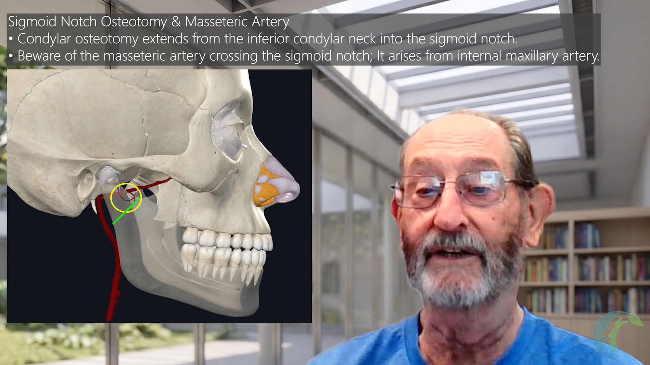
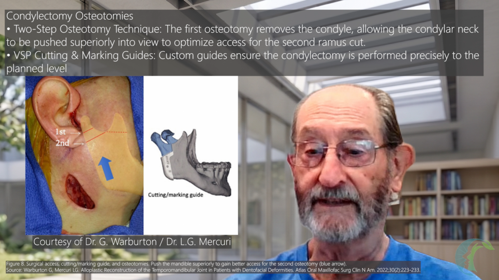
- When performing the condylectomy, the cut is typically made at the most inferior portion of the condylar process and extended into the sigmoid notch. Two-step osteotomy technique can help with exposure as the ramus will telescope into the preauricular incision for better exposure.
- Surgeons must be acutely aware of the masseteric artery located within the sigmoid notch. Accidental disruption of this artery during the osteotomy is a known complication that requires immediate intraoperative hemostasis.
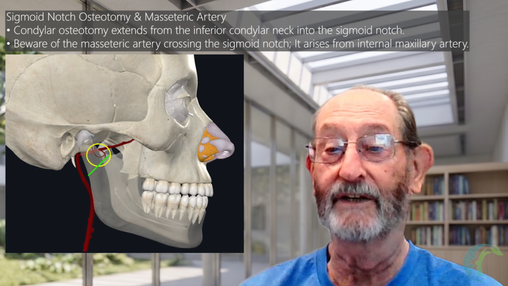
- To optimize exposure of the condyle through the preauricular incision, Dr. Mercuri typically recommends performing a two-osteotomy procedure. The first osteotomy and removal of the condylar head segment will allow the subcondylar region to telescope into the surgical field, allowing the more inferior osteotomy to be performed with improved exposure.
4.4 Drilling, Thermal Control, and Fixation Mechanics
- Drill Parameters: Use a low-speed, high-torque orthopedic drill operating between 250 RPM and 1,500 RPM.
- Thermal Necrosis Threshold: According to Dr. Mercuri, bone temperatures exceeding 42°C can cause thermal injury to osteocytes, potentially compromising osseointegration and contributing to early osteolysis and screw loosening.
- Drill Guide Mechanics: Dr. Mercuri recommends holding the drill guide perpendicular to the prosthesis screw holes to facilitate proper screw seating and avoid interference with the countersunk recess. Screw seats in the titanium ramus component feature machined countersunk recesses. Off-axis drilling forces the screw head to catch the rim of the recess during insertion, shearing or popping the screw head off.
- Screw Length Mechanics & Anatomic Risks:
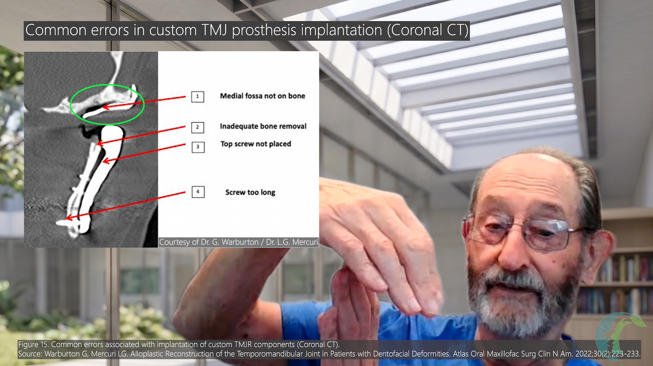
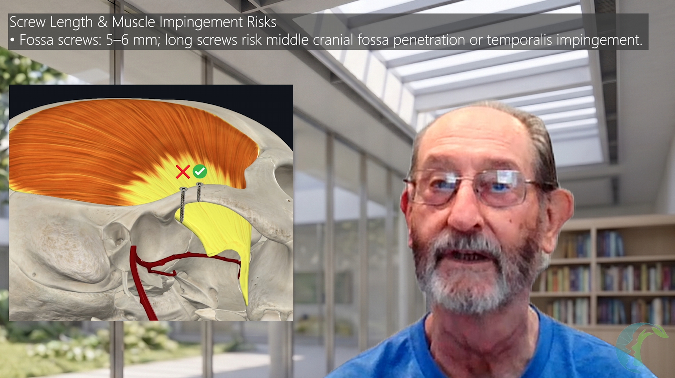
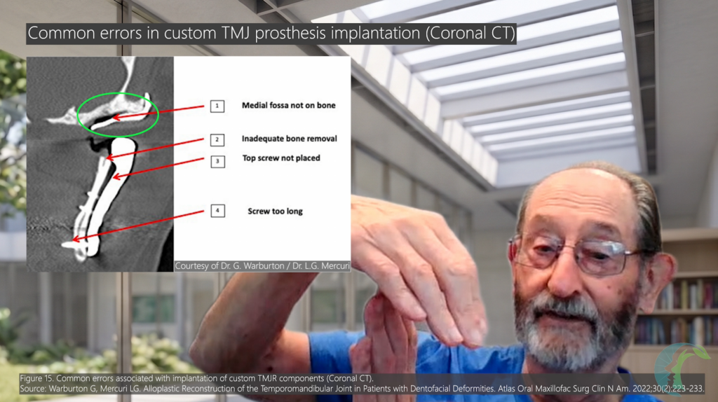
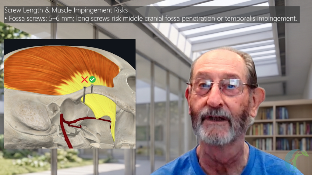
- Fossa Screws: Commonly 5–6 mm in this technique; final length should be determined by the patient-specific implant and available bone. Avoid excessively long screws as they may disrupt the middle cranial fossa and lead to a CSF leak from dural disruption.
- Ramus Screws: Commonly 10mm to 14mm; Must achieve bicortical engagement without disrupting the pterygoid muscle.
- Too Long Screws: Screws extending beyond the medial cortex impinge directly into the Medial Pterygoid Muscle or Temporalis Muscle tendon, causing persistent postoperative pain during mastication.
- Too Short: Fails to engage the medial cortex, predisposing to micro-motion and fixation failure.
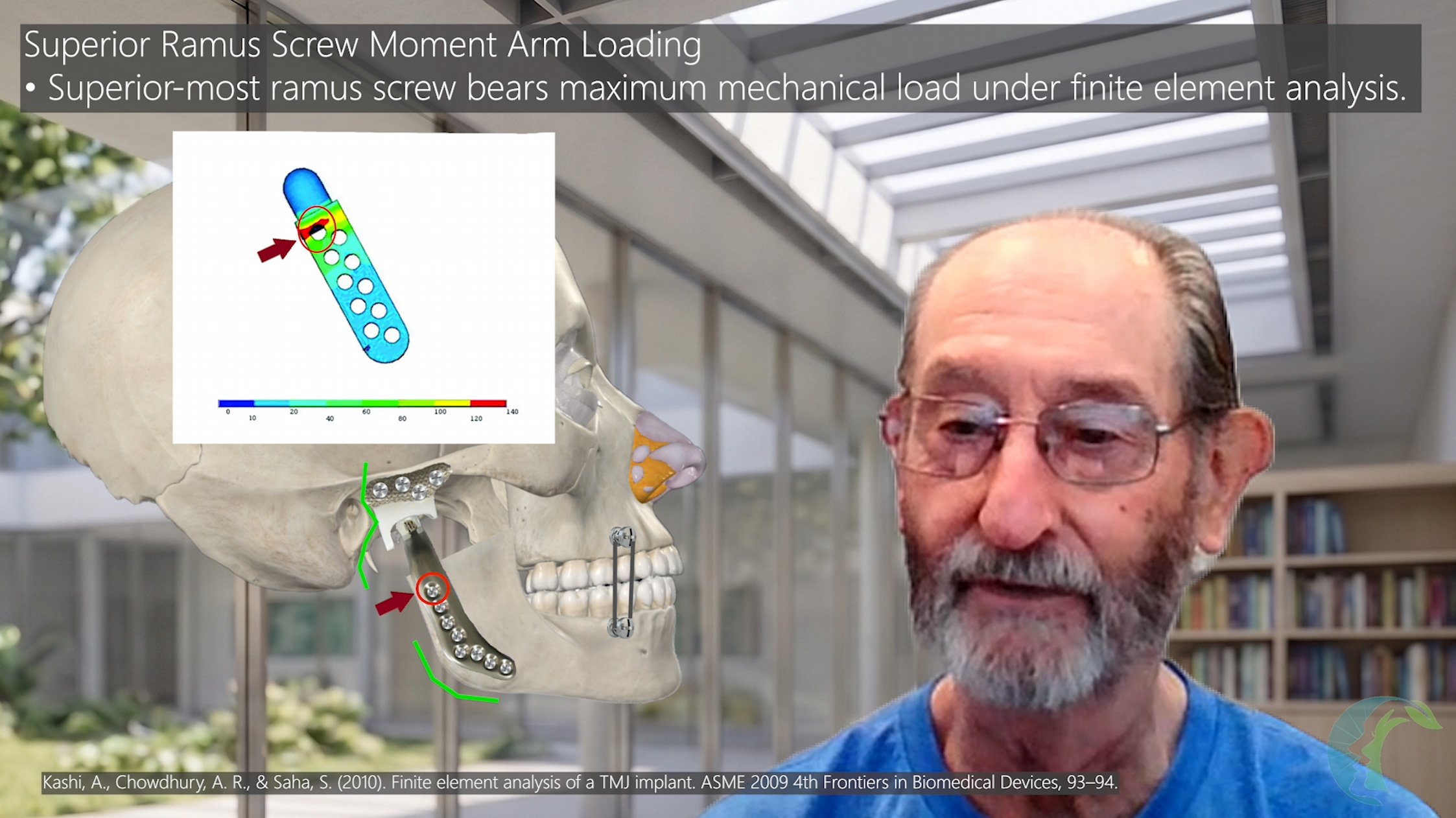
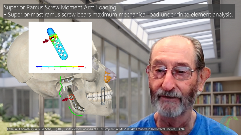
- Top Screw Priority: The most superior screw on the ramus plate handles the highest moment arm and mechanical stress loads under finite element analysis. Dr. Mercuri emphasizes achieving solid bicortical fixation at the most superior ramus screw, which experiences a high mechanical moment arm based on biomechanical considerations.
- Loose screws must be removed immediately and replaced with an emergency oversized screw; never leave a loose screw in situ.
- Complete Hole Utilization: Insert all manufactured screw holes. Do not omit screws based on partial fixation assumptions.
4.5 Autogenous Abdominal Fat Grafting
- Harvesting: Harvest a structural autogenous fat graft from the periumbilical region.
- Packing Technique: Pack fat densely around the joint space, filling all dead space surrounding the functional articular interface of the prosthesis. Use a blunt, smooth neurosurgical elevator or smooth instrument tip to avoid scratching the UHMWPE or metal surfaces.
- Benefits of Fat Grafting:
- Ossification Prevention: Dr. Mercuri recommends dense packing of autogenous abdominal fat around the joint space and residual dead space. This may prevent heterotopic ossification (HO), which may lead to ankylosis from abnormal bony fusion. HO is thought to arise, at least in part, from the surrounding traumatized or inflamed soft tissues, including the temporalis and medial pterygoid muscles, rather than exclusively from the joint space; packing fat may act as a physical barrier. (Mercuri & Saltzman, 2017).
- Dead Space Obliteration: Reduces residual dead space and may reduce hematoma formation.
- Hemostasis: Exhibits local hemostatic properties.
- Fibrous Metaplasia: Over time, the fat graft undergoes remodeling and may be replaced in part by fibrous connective tissue.
SECTION 5: POSTOPERATIVE MANAGEMENT, PHYSICAL THERAPY, AND REVISION SURGERY
5.1 Immediate Postoperative Care
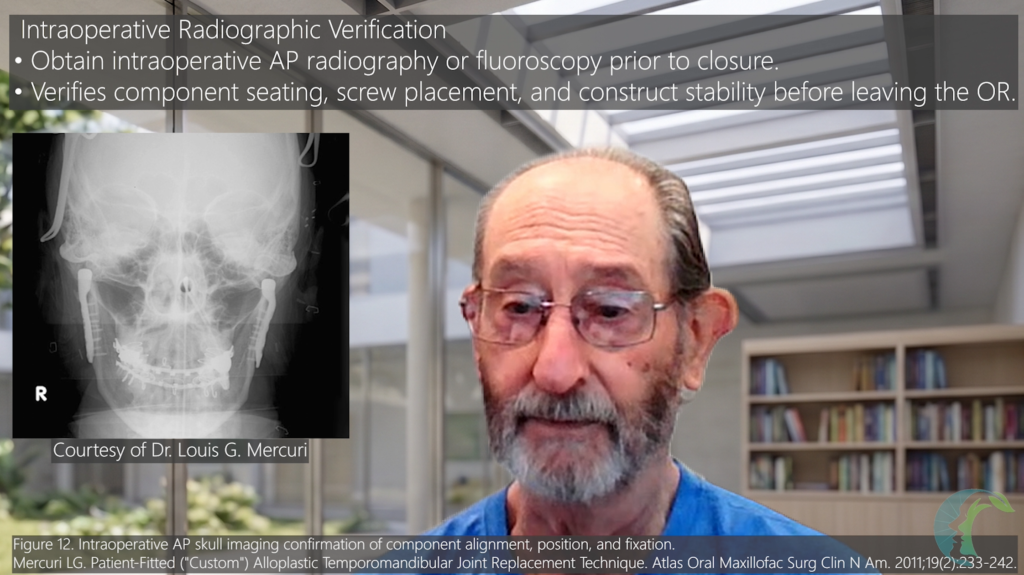
- Intraoperative Imaging Verification: Obtain Anteroposterior (AP) and lateral radiographs (or C-arm fluoroscopy) in the operating room prior to extubation. Verify component position, screw trajectory, condylar seating, and hardware integrity before leaving the operating room.
- Hemostasis & Wound Dressings:
- While a pressure dressing will adequately manage standard postoperative oozing, it will not solve an active bleeder. Prior to closure, surgeons must ensure all active bleeding has completely stopped and is reduced to no more than a gentle ooze.
- Place a compression dressing over the preauricular and transfacial surgical sites for 24 hours to prevent postoperative hematoma formation.
- Elastics & Maxillomandibular Fixation (MMF):
- Avoid routine post-op MMF or heavy elastics.
- Heavy MMF induces joint stiffness and secondary muscle conditioning errors.
- Exception: Light guiding elastics are indicated only if a concurrent coronoidectomy was performed, or to guide new occlusal settling in complex orthognathic/reconstructive cases.
5.2 Physical Rehabilitation Protocols
-
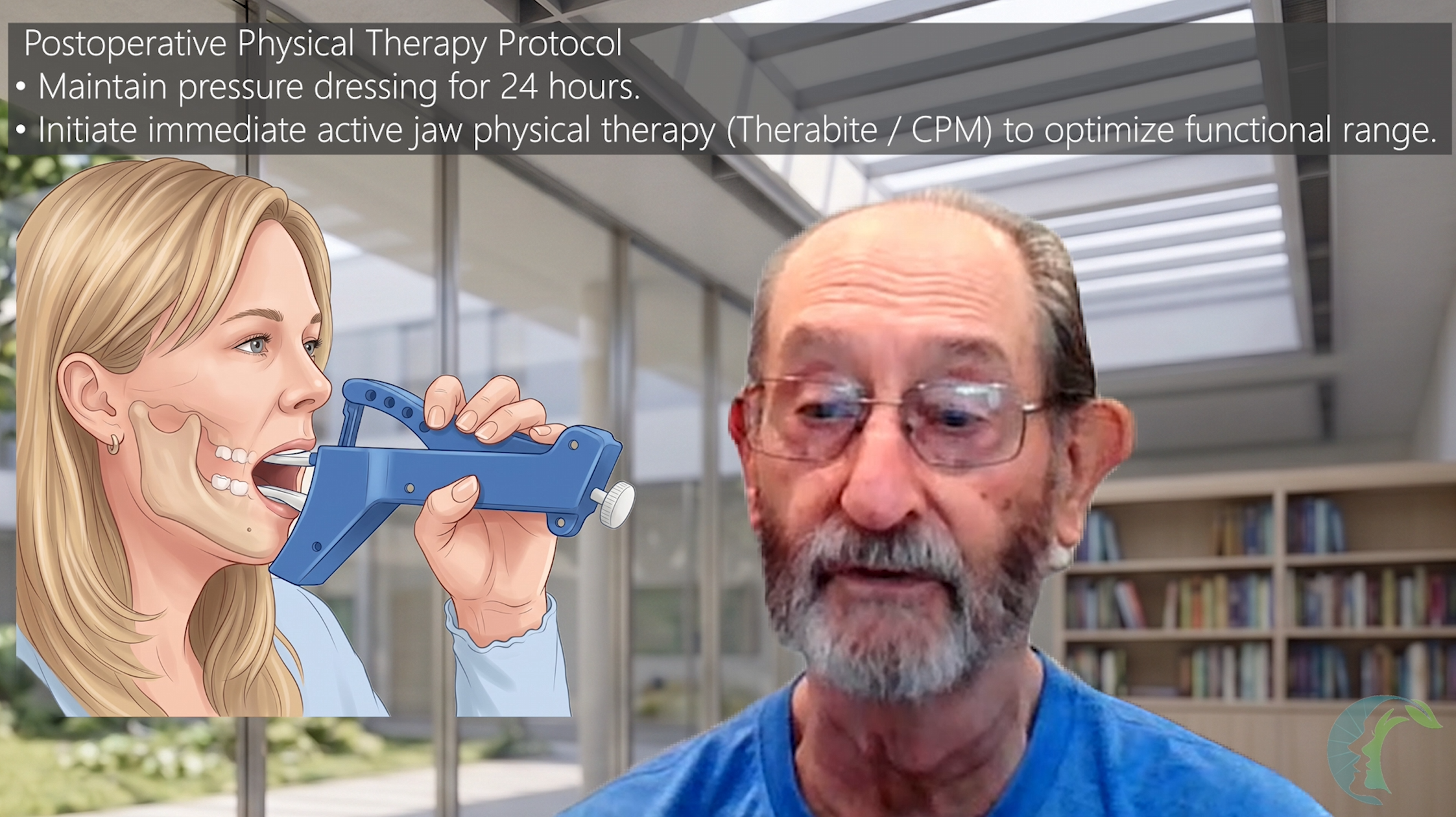
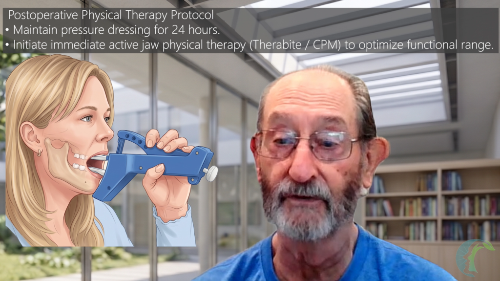
Early Mobilization: Dr. Mercuri recommends removal of the pressure dressing at approximately 24 hours, followed by immediate active range-of-motion (ROM) exercises using jaw mobilization devices (e.g., Therabite or mechanical jaw exercisers), provided there are no contraindications.
-
Continuous Passive Motion Rationale: Early mobilization is intended to promote functional remodeling and reduce peri-articular scar contracture.
-
Concomitant Le Fort I Osteotomies: If a simultaneous Le Fort I osteotomy was performed, delay aggressive mechanical jaw mobilization for 2 to 3 weeks to allow maxillary osseous healing.
5.3 Coronoid Process Management and Coronoidectomy
- Coronoidectomy vs. Coronoidotomy: In the setting of trismus, it is best to avoid coronoidotomy (osteotomy of the coronoid process). The temporalis muscle will pull the detached coronoid segment superiorly, where it scars and may reattach to the ramus, causing recurrent trismus within 1–2 years. Dr. Mercuri prefers a complete coronoidectomy rather than coronoidotomy in patients with significant trismus when coronoid intervention is indicated.
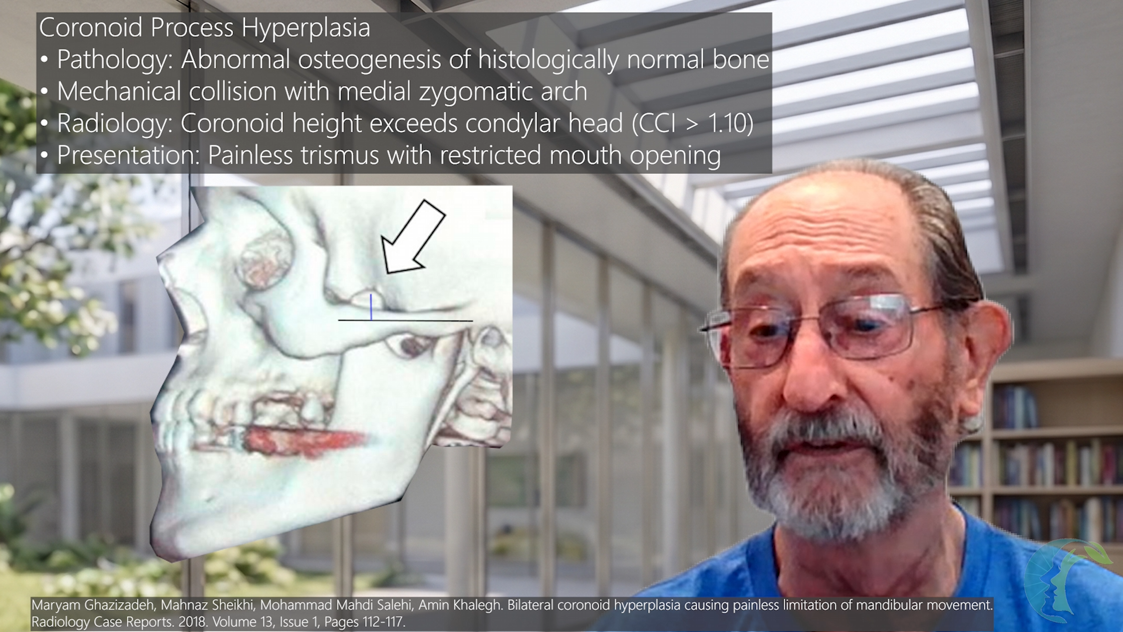
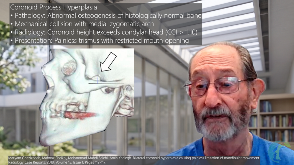
- Coronoidectomy is not routinely performed in typical TMJ replacement procedures, as it increases the potential for TMJ dislocation. However, if coronoid hyperplasia is present with a concern for trismus, a coronoidectomy is performed concurrently with TMJR.
SECTION 6: MASTER SURGICAL CHECKLIST
- Administer IV Cefazolin/Clindamycin 30 minutes or more prior to skin incision.
- Clip hair with electric clippers (NO razors).
- Rotate operating table 180 degrees; place anesthesia at foot of bed.
- Protect nares with dexamethasone cream; suture endotracheal tube to nasal septum.
- Place eye ointment, tape eyelids shut, and secure rigid goggles over eyes.
- Inspect EAC, place antibiotic/steroid-soaked cotton pledget into canal.
- Soak all prosthetic components in Povidone-Iodine solution prior to implantation.
- Through a preauricular incision, perform complete fossa soft tissue dissection down to bare bone while being careful to avoid injury to the middle meningeal artery.
- Dissect fossa medially until encountering the inferior dip (Foramen Spinosum boundary).
- Maintain smooth UHMWPE and CoCrMo surfaces; use non-metallic or plastic pushers.
- Drill screw holes using low-speed (250-1500 RPM) orthopedic drill with cold irrigation.
- Keep drill guide perpendicular to avoid shearing screw heads in countersunk recesses.
- Ensure superior ramus screw achieves solid bi-cortical engagement.
- Pack autogenous abdominal fat densely around the joint space and all dead space.
- Obtain intraoperative AP/Lateral radiographs or C-arm fluoroscopy prior to extubation.
- Perform postop otoscopy, micro-suction EAC, and remove all blood clots.
- Remove pressure dressing at 24 hours and initiate immediate active ROM exercises.
REFERENCES
- Mercuri LG, Wolford LM, Sanders B, White RD, Hurder A, Henderson W. Custom CAD/CAM total temporomandibular joint reconstruction system: preliminary multicenter report. J Oral Maxillofac Surg. 1995;53(2):106-15
- La Touche R, Paris-Alemany A, Hidalgo-Pérez A, et al. Evidence for Central Sensitization in Patients with Temporomandibular Disorders: A Systematic Review and Meta-analysis of Observational Studies. Pain Pract. 2018;18(3):388-409.
- Mercuri LG (Ed). Temporomandibular Joint Total Joint Replacement – TMJ TJR – A Comprehensive Reference for Researchers, Materials Scientists and Surgeons. Springer International Publishing. New York. 2016.
- Mercuri LG and Granquist EJ. End-Stage Temporomandibular Joint Disease. In, Miloro M, et al. (Eds.), Peterson’s Principles of Oral and Maxillofacial Surgery, 4th Springer Nature Switzerland AG. 2022. pp. 1706-1722.
- Mercuri LG (Ed). Indications for the Use of TMJ Replacement in Maxillofacial Surgery—an evidence-based review of the literature. Frontiers of Oral and Maxillofacial Medicine. AME Publishing Company Hong Kong. 2023.
- Mercuri LG (Ed). Temporomandibular Joint Total Joint Replacement (TMJR): A Comprehensive Reference for Surgeons, Clinicians, Materials Scientists, Biomedical Engineers, and Physical Therapists. Springer Nature. Switzerland AG. Forthcoming 2028.
- Mercuri LG, Abramowicz S. Temporomandibular Joint Arthritic Disease. In: Farah C, Balasubramaniam R, McCullough M, eds. Contemporary Oral Medicine. New York, NY: Springer International Publishing; 2018:1919-1954.
- Moss ML. The functional matrix hypothesis revisited. 1. The role of mechanotransduction. Am J Orthod Dentofacial Orthop. 1997 Jul;112(1):8-11. doi: 10.1016/s0889-5406(97)70267-1. (Cited in Section 1.2 – Skeletally Immature Patients)
- Mercuri LG. Microbial Biofilms – A Potential Source of Alloplastic Device Failure. J Oral Maxillofac Surg. 2006;64(8):1303-1309.
- McKenzie WS, Louis PJ. Temporomandibular total joint prosthesis infections: a ten-year retrospective analysis. Int J Oral Maxillofac Surg. 2017;46(5):596-602. doi:10.1016/j.ijom.2017.01.005. PMID 28161135.
- Chalmers PN, Beck L, Stertz I, Tashjian RZ. Hydrogen peroxide skin preparation reduces Cutibacterium acnes in shoulder arthroplasty: a prospective, blinded, controlled trial. J Shoulder Elbow Surg. 2019;28(8):1554-1561. doi:10.1016/j.jse.2019.03.038. PMID 31229329.
- Machinski M, et al. Chlorhexidine or Povidone-Iodine Solution Irrigation Versus Saline Irrigation for the Prevention of Postoperative Infections in Primary Total Joint Arthroplasty: A Systematic Review and Meta-Analysis. J Arthroplasty. 2025 Sep;40(9):2458-2466.e2
- Neto MQ, Radice S, Hall DJ, Mathew MT, Mercuri LG, Pourzal R. Alloys used in different temporomandibular joint reconstruction replacement prostheses exhibit variable microstructures and electrochemical properties.J Oral Maxillofac Surg. 2022;80(5):798-813.
- Mercuri LG. Patient-Fitted (“Custom”) Alloplastic Temporomandibular Joint Replacement Technique. In, Atlas of Oral and Maxillofacial Clinics of North America. Ness G. (Ed). Elsevier. Philadelphia. Atlas Oral Maxillofac Surg Clin North Am. 2011;19(2):233-242.
- Mercuri LG, Saltzman BMAcquired heterotopic ossification of the temporomandibular joint. Int J Oral Maxillofac Surg 2017;46(12):1562-1568.
- Mercuri LG. Alloplastic temporomandibular joint replacement: rationale for the use of custom devices. Int J Oral Maxillofac Surg. 2012;41(9):1033-1040.
- Wolford LM, Mercuri LG, Schneiderman ED, Movahed R, Allen W. Twenty-year follow-up study on a patient-fitted temporomandibular joint prosthesis: the Techmedica/TMJ Concepts device. J Oral Maxillofac Surg. 2015 May;73(5):952-960. doi: 10.1016/j.joms.2014.10.032.