
By Kathryn Bradburn MD, Nima Vahidi, MD, Derek Sheen MD, Thomas S. Lee MD
This is a case report on a trauma patient presenting with ZMC, NOE type 1, & Le Fort type 1 fractures with a lacrimal duct fracture, lateral canthus detachment and nasal airway obstruction. Preoperative imaging assessment and surgical planning process are discussed in detail.
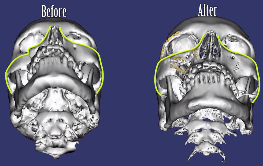
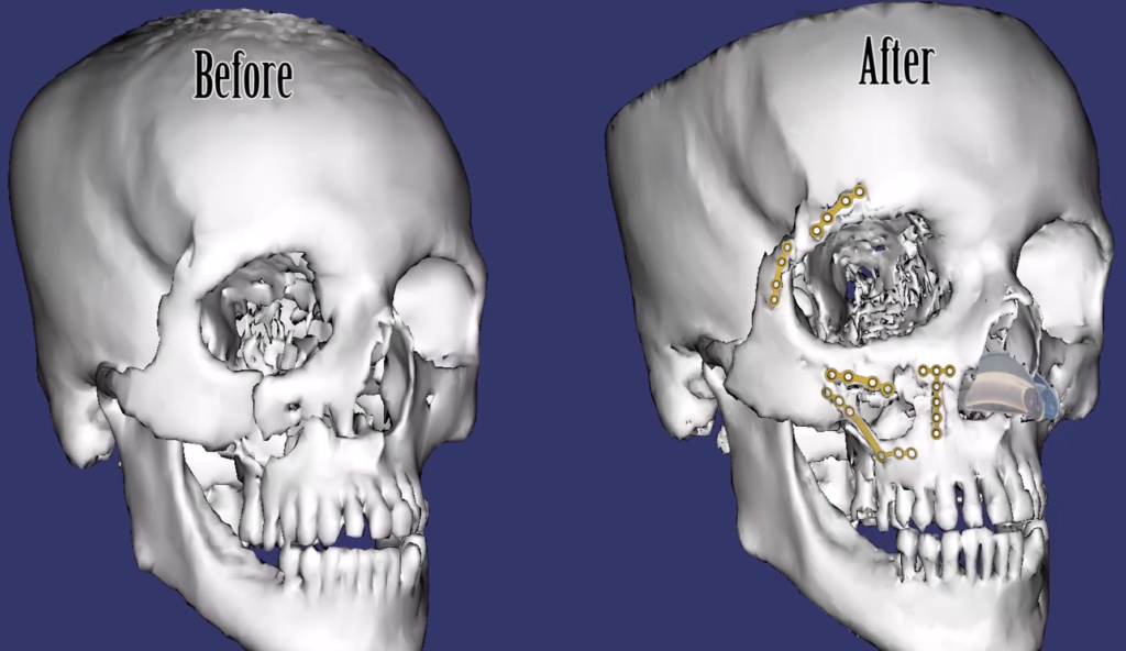
Identified Fractures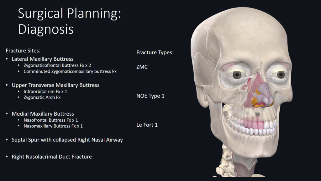
Surgical Indications & Goals 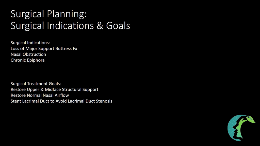
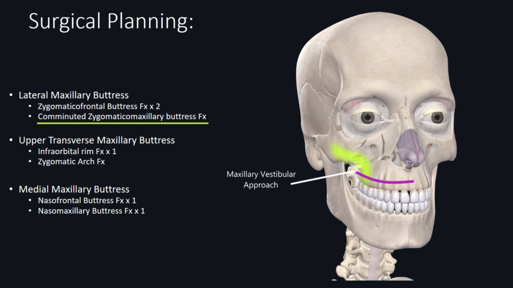
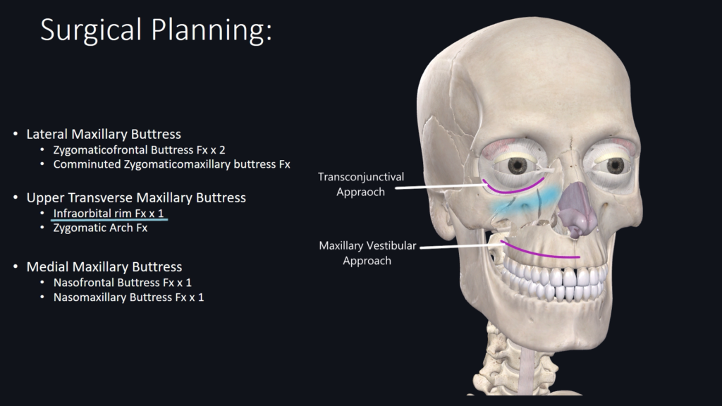
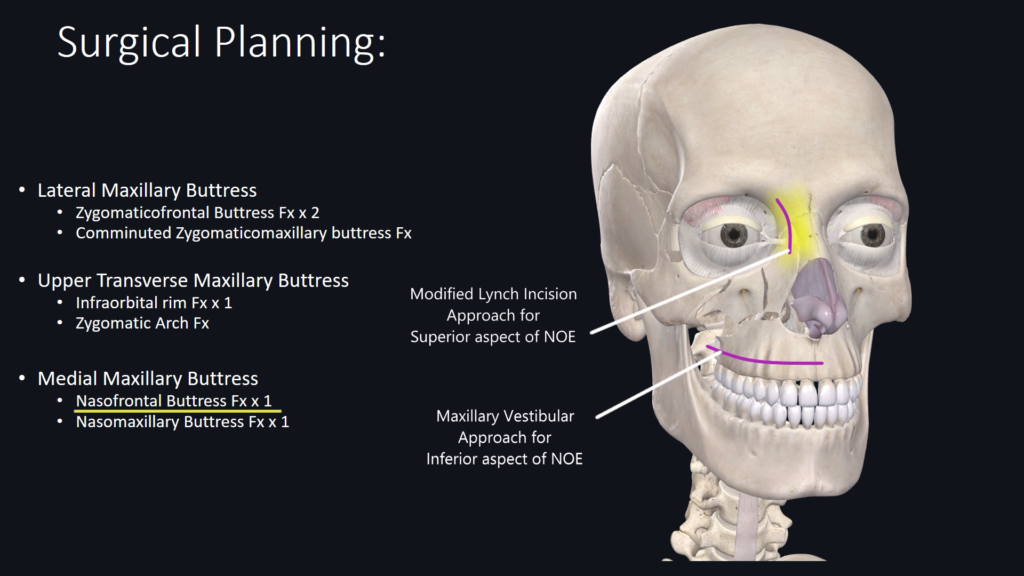
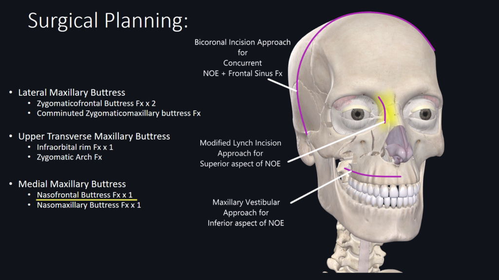
Surgical Approaches for Fracture Site 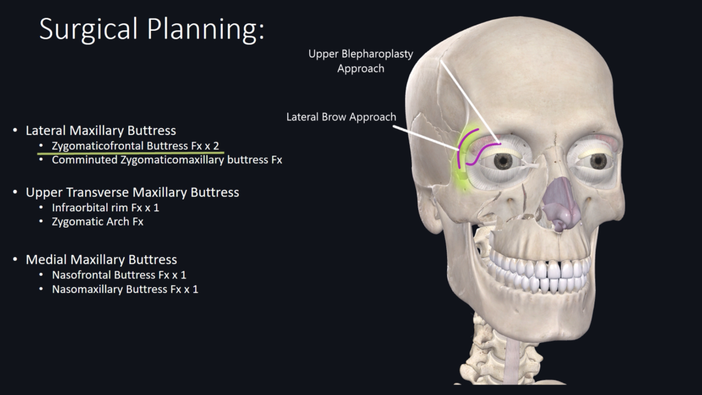
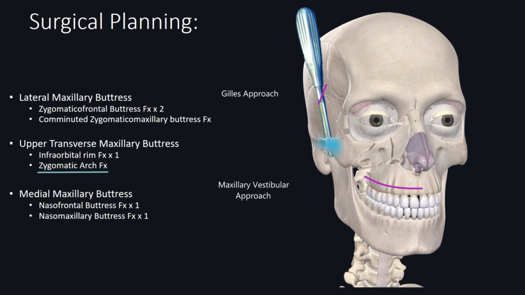
Sequence of Procedures 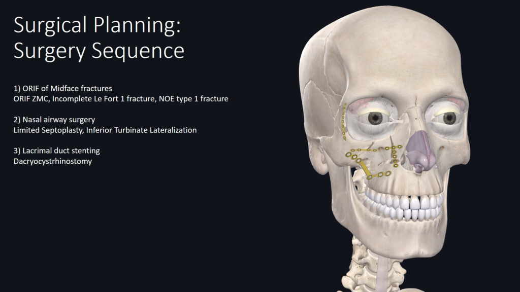
2 Responses
Hello Dr. Lee,
Great presentation.
Regarding nasolacrimal duct injury, could you please elaborate on the radiological findings that would prompt you to seriously consider dacryocystorhinostomy (DCR)?
In many cases, patients do not seem to develop epiphora during follow-up visits, and the literature also reports a relatively low incidence. Most recommendations suggest observation with follow-up and reserving DCR for patients who develop symptomatic epiphora.
I would appreciate your insights on when imaging findings might influence a more proactive approach.
Kindest regards
Dr. Siddhartha Rai
OMFS
Nepal
I agree that most lacrimal duct injury from NOE will not result in total obstruction. With that said, I think a lot of the issues are underreported because at least in USA, DCR is done by oculoplastic who does not routinely perform NOE fractures. So Facial fx get reparied by OMFS/ENT/Plastic surgery and get refer to oculoplastic when the pt develops lacrimal duct obstruction. ENT will perform endonasal approach with oculoplastic will perform external approach. So it is rare for one person to do the full spectrum from initial fx repair all the way to DCR. Also, most facial fx repair surgeons may not feel comfortable doing prophylactic stenting and to be honest, at the end of a long facial fx case, that is the last thing you want to deal with at the end of the case. I have a tendency to stent them more than others I think as I feel comfortable doing them and i can perform both external or internal approach as we don’t have oculoplastic coverage at my hospital. To answer your question, if I see on axial CT scan that lacrimal duct is completely disrupted, I will most stent it using crawford stent. If there is minimal displacement than I often will not stent it. I do agree that it is often very unpredictable who will develop the issue or not. personally, I think it is better to stent it than not if you can do the procedure safely and not laceration the duct in the process.